It’s a new year and many chiropractors are evaluating what will enhance their respective practices, particularly as it relates to their bottom line. One of the most common questions I get is: “Do I need to be credentialed to bill insurance, and what are the best plans to join?” It’s a loaded question – but one every DC ponders. Whether you're already in-network or pondering whether to join, here's what you need to know.
Diagnostic Ultrasound
This article is intended to be a brief discussion of the physics behind the imaging of diagnostic ultrasound. It may seem slightly tedious to discuss ultrasound in this manner, but I think that before we can appreciate this imaging modality, we need to know only superficially how the images are created - then we can examine some of the more clinical aspects of ultrasound.
As sound waves move through any medium, be it air or water, they are reflected by any solid objects. This is the basis of sonar. The sound wave moves through water and is reflected off a solid surface - for example, the bottom of the ocean. This reflected sound is termed an "echo." We know the speed of sound in water or air; we can calculate the distance to an obstacle or solid surface by measuring the time it takes for a pulse of sound to travel to the object and back again, then dividing by two; and we know the distance to the object or obstacle. (The formula: distance = speed x time. This is the total distance traveled by the sound wave. Divide this by two to calculate the one-way distance, or the distance from the source of the sound to the obstacle. This is basically the way sonar navigation works.)
High-frequency sound pulses are used because they travel further without being absorbed. Ultrasonic frequencies are all above 20kHz. Ultrasonic frequencies can be used to detect the topography of the surface of the ocean; the depth of a shoal of fish; or flaws in a piece of metal. Hand-held instruments can even utilize ultrasonic frequencies to measure the size of a room. Remember: The speed of sound varies from one medium to another, and also depends on temperature, pressure and other factors, so it's not a simple arithmetic equation; however, if all the factors are known, it's only a matter of calculating.
"So, where is the image?" you ask. Be patient - we are not quite there yet. So far, we've only discussed distance to objects or distance to changes in solid objects with the use of sound. I want to touch on the production, detection, transmission and reflection of the ultrasound. (Hang in there: This is interesting, partly because we use some of this in our daily practice, even though we probably don't think much about it. [Remember the term "piezoelectric?"] Some of us still use ultrasound for treating musculoskeletal problems.)
Some materials contract or expand when placed in an electrical field. If the electrical field is reversed, the material will again expand or contract, depending on what state it was initially in. This procedure can be done continuously; the electrical field can be alternated rapidly, causing the material to expand or contract at such a high rate that it vibrates. This vibration, largest when the electrical field stimulates at the natural frequency of the material, is an example of resonance. The vibrations are passed through any adjacent material as a longitudinal wave, producing the sound wave. A number of materials behave in this manner, the most common being quartz crystals. The substance most commonly used to produce the ultrasound wave is a synthetic ceramic lead zirconate titanate crystal. This crystal is sliced into thickness that is half a wavelength of the desired ultrasound frequency, since this thickness will be the fundamental frequency the crystal will produce.
So, where is the image, already? Hold on - we have only discussed how we produce the ultrasound. Now, we need to detect the ultrasound, so we can obtain the data for creating the image.
To detect ultrasound, we again use the piezoelectric effect, because it also works in reverse. If one crystal is being vibrated or resonated by an alternating electrical field, a second crystal can respond to any returning ultrasound from the first crystal. This returning ultrasound will cause the second crystal to vibrate, producing an alternating electrical field that can be amplified and processed in several ways (which I will not discuss in this article because, admittedly, I don't have the expertise to delve deeper into physics).
Thus far, we have described crystals that utilize the piezoelectric effect to transmit and receive ultrasound. Generally, these crystals are built into the same handheld unit: the transducer.
Now, we can finally discuss how we obtain the ultrasound image! Here is the basic process:
When sound travels through a medium and hits a boundary with another medium, some of the sound will pass through the second medium, and some will be reflected by that difference (or boundary) between the two different mediums. The same thing happens when light is reflected by glass or water: Some of the light will pass through the transparent element; some will be reflected back. The exact percentage transmitted or reflected depends on the difference between the two materials. With ultrasound, this difference is gauged by the "acoustic impedance" of the material. The greater the difference in impedance, the more sound will be reflected back, rather than transmitted through. The following table shows some of the more common impedances.
Medium | Impedance (standard units) |
air | 0.000429 |
water | 1.50 |
blood | 1.59 |
fat | 1.38 |
muscle | 1.70 |
bone (avg.) | 6.5 |
soft tissue | 1.63 |
brain | 1.58 |
As you can see, air, water and bone have quite different impedances. This means that a beam of ultrasound hitting the water from air will be almost entirely reflected away. This is the case when a beam of ultrasound hits skin from air. Because of this, we need to use a "coupling medium" for the ultrasound to be transmitted through the skin. So, a coupling agent is used between the patient's skin and the probe or transducer. Note, however, that different body layers, such as fat, muscle and soft tissue have similar impedances, so most of the beam of ultrasound will pass from layer to layer with only a small amount of reflection. This is how we produce the image through ultrasound. Only a small difference in impedance is necessary to obtain a good resolution of the interface between two layers, about 1 percent. Initially, ultrasound signals were evaluated in this manner. The reflections from the different layers of tissues were reflected back at different time intervals and amplitudes, depending on their impedance to the ultrasound and the distance from its origin. So, what you get is a graph in which the x-axis is the amplitude, and the y-axis is the time and/or distance. This is called an "A-scan," which is difficult to interpret, and currently is used only for measuring ranges or the thickness of a body organ (the eye, in particular).
The A-scan has been replaced mainly by the "B-scan." With this method, the ultrasound signal is not simply displayed as a graph, but as an image. The amplitude of the signal controls the brightness of an area on a screen - producing the "B" in B-scan. A single pulse of ultrasound passing through layers of different tissues will give a series of focal areas of brightness corresponding to the amplitude of the reflected pulse of the different layers of tissue. The largest amplitude will give the greatest brightness; the smallest will provide the darkest.
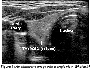
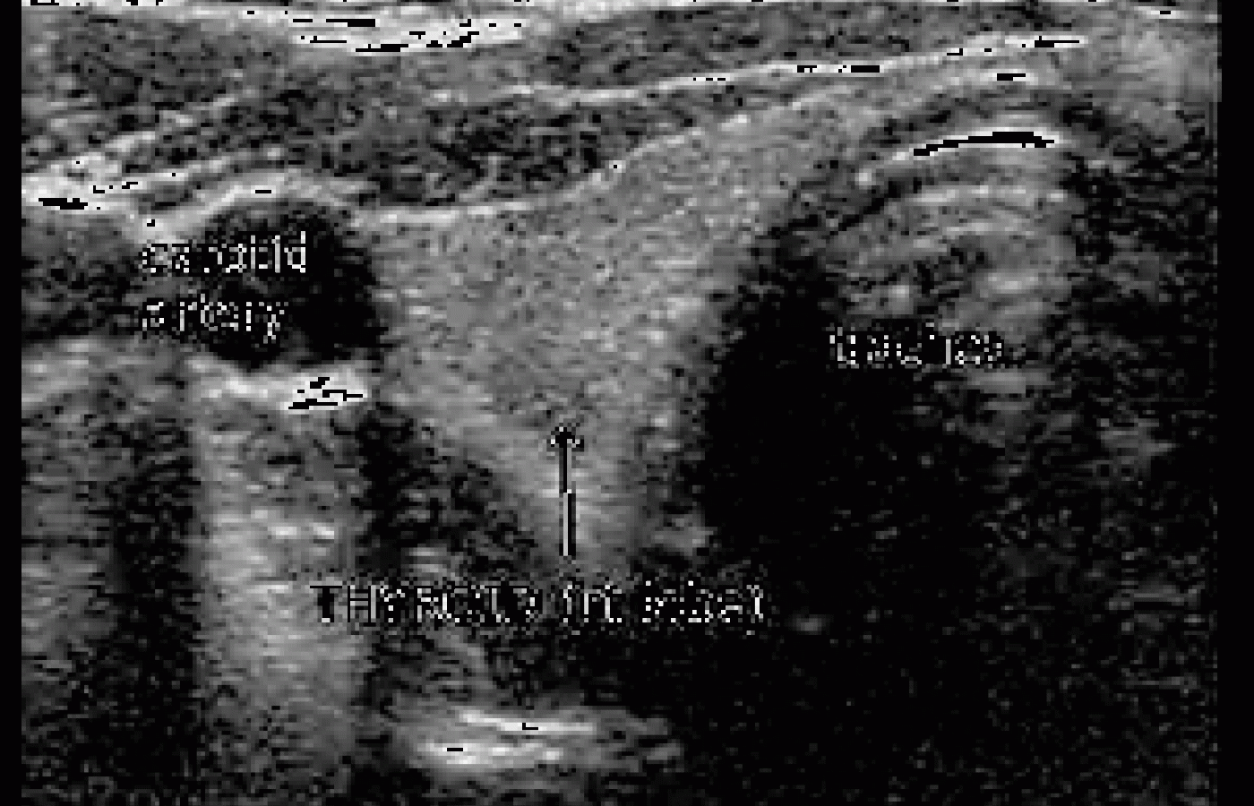
A 2-D image is constructed by "rocking" the probe from side to side, which alters the angle of the beam, giving a slightly different view of a region. At the same time, the probe is moved horizontally across the body. This is not a technique one can acquire in a few minutes; it takes some practice to obtain a diagnostic image. A significant amount of data needs to be processed to produce an image. As you can imagine, the image can change drastically, depending on the technical ability of the person manipulating the probe. Imaging technology is improving, but it still takes a skilled specialist to interpret the findings. In Figure 1, without knowing the location of this image and how it was obtained, it is just a grayscale study; what it is exactly is anyone's guess.
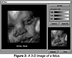
At this point in our discussion, it is not important to know what part of the anatomy this image represents, but suffice to say, it is not an imaging modality easily comprehended by the untrained eye. However, there are more sophisticated versions of 2-D B-scans, including real-time B-scans and 3-D imaging, which make viewing and evaluating the anatomy much easier. (see Figure 2.)


Real-time B-scans allow body structures to be evaluated while moving. This makes ultrasound images useful in diagnosing tendon tears, bleeding or other fluid collections within the muscles, bursae and joints to be detected. Unfortunately, to date, ultrasound has not been proven useful in detecting whiplash injuries or other spinal problems in adults. However, there is room here for further investigation, particularly considering the rapid advances in technology.
To complete this brief review on ultrasound, I must mention Doppler methods and ultrasound therapy. When ultrasound is reflected from a moving surface, the frequency of the sound is altered slightly, depending on the speed of the movement of the surface. This is the Doppler effect, used in ultrasound imaging to examine the movement of fluids (such as the flow of blood in arteries and veins), allowing the location of blockages to be determined precisely. In recent years, Doppler ultrasound scanners have been able to display an estimate of blood velocity in real time. Several different velocity-estimation systems are available. Color-flow mapping systems interrogate the whole region of the body and show a real-time image of velocity, which is useful in diagnosing heart valve problems, stenosis of veins and arteries, and other hemodynamic problems.
The use of ultrasound as therapy is a final, but no less important, area of discussion. Intense beams of ultrasound are used presently to heat tissues inside the body. It has been used effectively to break up gallstones and kidney stones, and to relieve the symptoms of arthritis. The exact mechanism of this modality is not completely understood and is beyond the scope of this article.
In summary, medical ultrasound is still an evolving imaging modality. It has many advantages when compared with other modalities:
- No apparent harmful effects have been detected at the intensity levels used today, compared with X-rays, CT scans and radioactive isotope studies. Ultrasound uses no ionizing radiation.
- It is noninvasive and usually painless, but also can be used in conjunction with invasive procedures (i.e., needle biopsies and aspiration of fluid).
- It does not affect cardiac pacemakers, ferromagnetic implants or fragments in the body, as does the strong magnetic field of an MR scan.
- It is inexpensive, fast and convenient compared to techniques such as X-rays, CT, and MR scans. The equipment is portable and can be utilized even when the patient is not ambulatory.
- It is particularly suited to imaging soft tissues, such as the heart, liver and kidneys, and examining blood vessels and blood flow.
- The real-time feature of ultrasound is relatively unique, which not only allows for the evaluation of blood flow, but also can be used to evaluate the motion of tendons in large joints (i.e., the shoulder). The possible applications are expanding.
Of course, the limitations of ultrasound imaging are important considerations; as with any imaging modality, the limitations are due to the physics involved in acquiring the images.
- Because ultrasound is based on waves reflected by air or gas, it is not an imaging modality that can be used to examine the bowel.
- Ultrasound has difficulty penetrating bone; therefore, it can only demonstrate the very outer surface of the bony structures, not what lies within or beyond. CT and MR are far better modalities when it comes to evaluating osseous and soft-tissue structures around osseous structures (e.g., the spine).
- Ultrasound resolution is still limited, and there are many situations in which even X-rays produce a more diagnostic image.
- The interpretation of ultrasound images requires highly skilled specialists, especially for complicated procedures.
Please keep in mind this is an extremely brief review of the more common uses of ultrasound. For more detailed information regarding its clinical uses, review the specific topics in the medical literature currently available online and elsewhere.
(As in the past, I thank Philips Medical Systems - online at www4.medical.philips.com:80/Education/education.asp - for permission to use its images.)
Deborah Pate, DC, DACBR
San Diego, California
patedacbr@cox.net


