New York's highest court of appeals has held that no-fault insurers cannot deny no-fault benefits where they unilaterally determine that a provider has committed misconduct based upon alleged fraudulent conduct. The Court held that this authority belongs solely to state regulators, specifically New York's Board of Regents, which oversees professional licensing and discipline. This follows a similar recent ruling in Florida reported in this publication.
Common Intrapelvic Soft Tissue Calcification
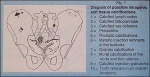
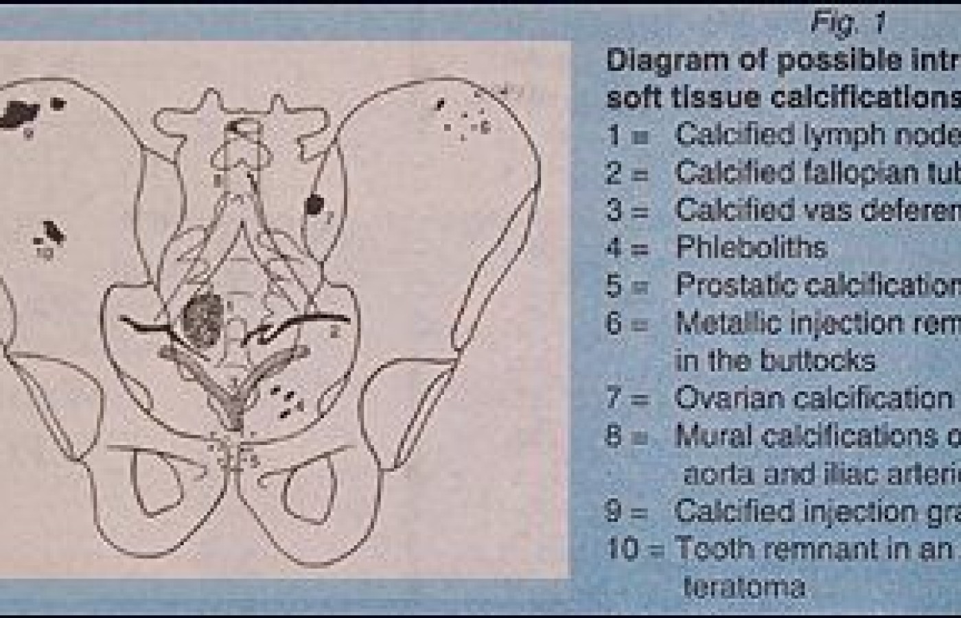
Intrapelvic calcifications are fairly common findings on AP lumbar views. This diagram lists some of the more frequent causes of calcification.
- Phleboliths are very common and are a calcified thrombus within a vein. Most adults will have a few in the pelvic basin.
- Arterial calcification in the large arteries such as the aorta and common iliac vessels represents the most common form of arterial calcification atherosclerosis.
- Subcutaneous and intramuscular injections can cause fat necrosis which will also calcify, often seen in the gluteal region.
- Peripheral mesenteric lymph nodes commonly will calcify after being involved in a previous infection.
- Calcification can also be found in the prostate caused by renal calculi.
- The vas deferens may calcify, but is usually found only in men with long standing diabetes.
- Calcification is often found in uterine fibroids.
- Calcification is frequently present in benign and malignant ovarian tumors.
- Most calcifications found in the pelvis are relatively benign, however, if there is a concern that the calcification may involve a neoplastic process, a second opinion should be obtained. Many patients do not have significant clinical symptoms early on, therefore, the AP lumbar view may be the initial study that reveals the truly significant pathological process.

Deborah Pate, DC, DACBR
San Diego, California


