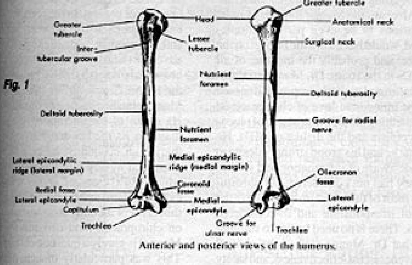
There are several common radiographic findings, seen in the humerus that simulate a pathological process. I'd like to review a few of them as they are relatively common findings.
In infants and even adolescents there is often a double contour seen along the lateral or medial metaphyseal region of the humerus, which is a normal finding (Fig. 2). In adults we often see a vertically oriented radiolucent line along the humeral shaft, that is a vascular channel, and may be mistaken for a fracture line. Commonly seen in adults is a radiolucency along the humeral shaft, caused by overlying muscles or soft tissues of the lateral chest wall such as voluminous breats (Fig. 3).
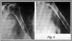
One of the most common questions I am asked is regarding the deltoid tubercle of the humerus. This tubercle is the region of insertion of the deltoid muscle onto the humerus. Generally it is not well visualized, however in some persons it can be very prominent. It generally appears as an irregularity in the contour of the cortex at the lateral proximal aspect of the humeral shaft (Fig. 4). At times however, this area can appear bubbly or cystic, with thickening of the cortex giving the humerus an expanded appearance. Frequently, a strange web-like pattern of sclerotic trabecular densities can be seen just underneath the cortical margin (Fig. 5). Similar changes are found at the insertion of the major pectoralis muscle along the crest of the major tubercle, slightly more proximal to the deltoid tubercle (Fig. 6).
Before alarming the patient, evaluate the humerus with these common radiographic findings in mind. If you're uncertain have a specialist review the study.
Recommended References:
Keats, Atlas of Normal Roentgen Variants.
Kohler/Zimmer, Borderlands of Normal and Early Pathologic Findings in Skeletal Radiography.
Building on a historic March 2026 meeting between Make America Healthy Again and chiropractic leadership, MAHA has announced the launch of the MAHA Chiropractic Hub, “a coordinated national partnership uniting MAHA Center, MAHA Action, and the chiropractic profession, including national associations, state organizations, practitioners, educators, researchers, and patient advocates. The Chiropractic Hub will advance federal policy, expand patient access, and build broad public support for chiropractic care across America.”
The chiropractic profession is confronting one of the most significant federal regulatory threats it has faced in decades. A new U.S. Department of Education (ED) accountability framework – commonly referred to as the “Do No Harm” (DNH) regulation – could place many chiropractic programs at risk of losing access to federal financial aid (student loans), potentially reshaping the future of chiropractic education and workforce development across the United States.
Pain has become the dominant language of musculoskeletal healthcare. Numeric pain-rating scales and symptom reports are routinely used as primary indicators of clinical success. But while pain reduction is meaningful, it is an incomplete and often misleading representation of recovery. This has real consequences for patient adherence, long-term outcomes, and how conservative care is perceived within the broader healthcare system.