With subscapularis syndrome introduced as an upper-extremity analog to piriformis syndrome, the next diagnostic question becomes essential: Why does the subscapularis become dysfunctional in the first place? Identifying the muscle alone is insufficient for durable clinical outcomes.
Instead, clinicians must understand the upstream biomechanical and neuromuscular factors that create a compensatory environment in which subscapularis dysfunction emerges as a primary driver of symptoms.
Scapular dyskinesis – defined as altered scapular position, motion or timing – represents the most common and clinically relevant upstream contributor to subscapularis syndrome. When scapular mechanics are disrupted, normal force coupling at the glenohumeral joint is compromised, placing excessive stabilizing demand on the subscapularis.
Over time, this altered loading environment transforms an adaptive stabilizer into a persistent pain generator capable of producing pseudo-radicular symptoms.
The Scapula’s Role in Shoulder Function
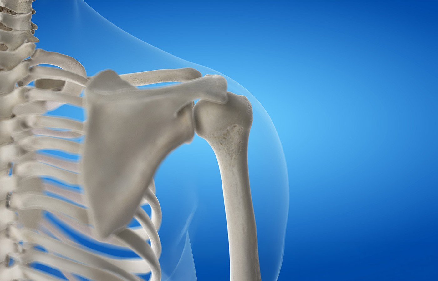
The scapula serves as the foundational platform for upper-extremity movement, linking the trunk to the arm and providing a stable base for force transfer. Efficient shoulder function depends on coordinated scapulothoracic and glenohumeral motion, particularly during overhead activity. When this coordination is disrupted, the burden of maintaining joint stability shifts to the rotator cuff – most notably the subscapularis.
Scapular dyskinesis may present as excessive anterior tilt, internal rotation, reduced upward rotation, or delayed activation during arm elevation. These alterations compromise the normal length-tension relationships of the rotator cuff and increase the demand placed on muscles responsible for anterior stabilization. The subscapularis, due to its size, depth and tonic role, often assumes this compensatory load.
From Compensation to Dysfunction
Initially, increased subscapularis activation functions as a protective mechanism, enhancing joint stability in the presence of scapular control deficits. However, when this demand becomes chronic, the muscle transitions from stabilizer to stress bearer. Sustained hypertonicity, reduced excursion, and altered recruitment patterns contribute to myofascial trigger-point development and impaired neuromuscular control.
This process mirrors mechanisms observed in piriformis syndrome, where altered pelvic or hip mechanics increase tonic demand on the piriformis, leading to distal symptom referral. In both cases, the muscle’s stabilizing role becomes maladaptive, generating pain patterns that extend beyond the local region and mimic neural pathology.
Clinical Observations in High-Demand Populations
The relationship between scapular dyskinesis and subscapularis dysfunction becomes particularly evident in high-demand populations, such as overhead athletes. Clinical experience working with professional baseball pitchers highlights this pattern clearly. Many athletes presented with diffuse arm pain, perceived weakness or neural-type symptoms despite minimal cervical findings and inconsistent neurological examination results.
In these cases, scapular control deficits were consistently present. Altered scapular timing during the throwing cycle increased reliance on the subscapularis for anterior stability during late cocking and deceleration phases. Addressing cervical findings alone rarely produced lasting improvement. However, when scapular mechanics and subscapularis function were restored, symptoms often resolved without escalation to invasive spine-directed care.
While elite athletes represent an extreme example, the underlying mechanisms are not exclusive to high-level sport. Desk-bound postures, repetitive upper-extremity use, prior shoulder injury, and deconditioning commonly produce similar scapular control deficits in the general population. The difference lies in magnitude, not mechanism.
Why Cervical Findings Can Be Misleading
Scapular dyskinesis frequently coexists with cervical joint dysfunction, postural abnormalities or degenerative disc changes. This coexistence can obscure the true driver of symptoms, particularly when imaging reveals structural changes that appear to justify a spinal diagnosis. However, cervical findings may represent secondary adaptations rather than primary pain generators.
When scapular dyskinesis alters shoulder mechanics, compensatory cervical muscle activation often follows. This secondary cervical involvement can produce local discomfort and positive orthopedic findings without being the dominant source of distal symptoms. Treating cervical findings in isolation may reduce local pain temporarily while leaving the primary scapular / subscapularis dysfunction unaddressed.
Implications for Clinical Reasoning
Recognizing scapular dyskinesis as an upstream driver reframes subscapularis syndrome from a localized muscle issue to a regional control problem. This perspective is critical for clinical reasoning and case management. Without addressing scapular mechanics, interventions targeting the subscapularis alone may provide incomplete or transient relief.
Conversely, identifying and correcting scapular dyskinesis reduces compensatory stabilizing demand on the subscapularis, allowing tissue tone, neuromuscular control, and afferent signaling to normalize. This approach aligns with the principles of myogenic pseudo-radiculopathy, emphasizing functional drivers over structural assumptions.
Preparing for Differential Diagnosis
Understanding the scapular–subscapularis relationship also sharpens differential diagnosis. When upper-extremity symptoms are reproduced by scapular loading, fatigue or repetitive shoulder activity – and when neurological findings remain inconsistent – clinicians should consider scapular dyskinesis–driven subscapularis syndrome as a primary contributor. This distinction is essential before escalating care toward invasive spinal procedures.
What’s Next...
The next installment in this series will translate these concepts into practical application, outlining a structured approach to differentiating subscapularis-driven pseudo-radiculopathy from true cervical radiculopathy in daily clinical practice.
Editor’s Note: For previous installments in this seven-part series (parts 1-4), click here.
Resources
- Kibler WB, McMullen J. Scapular dyskinesis and its relation to shoulder pain. J Am Acad Orthop Surg, 2003;11(2):142-151.
- Kibler WB, Sciascia A, Wilkes T. Scapular dyskinesis and its relation to shoulder injury. J Am Acad Orthop Surg, 2012;20(6):364-372.
- McClure P, et al. Shoulder function and 3-dimensional scapular kinematics in people with and without shoulder impingement syndrome. Phys Ther, 2006;86(8):1075-1090.
- Cools AM, et al. Rehabilitation of scapular dyskinesis: from the office worker to the elite overhead athlete. Br J Sports Med, 2014;48(8):692-697.
- Lewis JS. Rotator cuff related shoulder pain: assessment, management and uncertainties. Manual Therapy, 2016;23:57-68.
- Woolf CJ. Central sensitization: implications for the diagnosis and treatment of pain. Pain, 2011;152(3 Suppl):S2-S15.