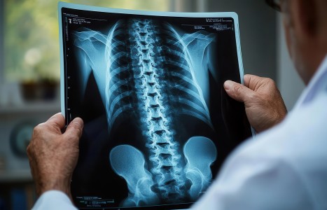
With the concepts of myogenic pseudo-radiculopathy, subscapularis syndrome and scapular dyskinesis established, the clinician’s next responsibility is practical differentiation. The distinction between subscapularis-driven pseudo-radiculopathy and true cervical radiculopathy is not academic; it directly determines case management, treatment escalation, and patient risk exposure.
While cervical disc pathology can unquestionably produce radiculopathy, many patients presenting with arm pain, paresthesia or perceived weakness do not exhibit the clinical behavior of true nerve-root compromise. In these cases, failure to differentiate the primary pain generator may lead to unnecessary spinal injections, nerve ablation procedures or surgical consultation despite limited likelihood of benefit.
Understanding True Cervical Radiculopathy
True cervical radiculopathy is defined by irritation or compression of a cervical nerve root and is typically characterized by a recognizable constellation of findings. Symptoms tend to follow a reproducible dermatomal distribution and are accompanied by objective neurological changes, including consistent sensory loss, myotomal weakness, reflex changes, or progressive neurological deficit.
In these cases, symptom reproduction is often closely linked to cervical spine loading, positioning or foraminal compression. Imaging findings that correlate anatomically and clinically strengthen diagnostic confidence. When these criteria are met, escalation to interventional or surgical care may be appropriate following a conservative trial.
However, this presentation represents a subset – not the majority – of patients labeled with “cervical radiculopathy” in daily practice.
Clinical Features Suggestive of Subscapularis Syndrome
Subscapularis-driven pseudo-radiculopathy presents differently. Rather than clean dermatomal patterns, symptoms are typically diffuse, variable and poorly localized. Patients may report deep shoulder pain, vague arm discomfort, fatigue, or intermittent paresthesia that does not consistently map to a single nerve root.
Neurological examination in these cases is often non-contributory. Reflexes remain intact, sensory changes are inconsistent or non-reproducible, and strength deficits – if present – are more reflective of inhibition than true myotomal loss. Importantly, symptoms may fluctuate with shoulder loading, scapular position, or repetitive upper-extremity activity rather than isolated cervical movement.
When such findings are present, clinicians should resist defaulting to a spinal diagnosis based solely on imaging abnormalities.
The Role of Imaging in Differential Diagnosis
Cervical imaging plays an important but limited role in differential diagnosis. Disc bulges, degenerative changes and foraminal narrowing are highly prevalent in asymptomatic populations. Their presence does not establish causality. In cases of subscapularis syndrome, imaging findings often coexist but function as secondary or incidental contributors rather than primary pain generators.
A key diagnostic error occurs when structural findings are interpreted as the dominant cause despite symptom behavior that does not align with nerve-root pathology. Imaging should be used to support clinical reasoning, not replace it.
Movement and Load-Based Differentiation
One of the most reliable methods for distinguishing subscapularis syndrome from cervical radiculopathy is symptom response to movement and load. In true radiculopathy, symptoms are typically reproduced with cervical spine provocation and relieved with decompression. In contrast, subscapularis-driven symptoms are more likely to be influenced by shoulder position, scapular loading, resisted internal rotation, or prolonged upper-extremity activity.
Fatigue-based symptom reproduction is particularly telling. Patients with subscapularis syndrome often report worsening symptoms with repetitive use or sustained postures rather than immediate exacerbation with cervical movement. This pattern reflects muscular endurance failure and neuromuscular control deficits rather than neural compression.
Primary vs. Secondary Pain Generators Revisited
Differential diagnosis hinges on identifying the primary driver of symptoms. Cervical joint dysfunction, postural abnormalities or degenerative changes may contribute to the overall clinical picture, but they do not necessarily represent the dominant pain source. In subscapularis syndrome, scapular dyskinesis and compensatory subscapularis overload often serve as the primary generator, while cervical findings represent secondary adaptations.
This hierarchy is critical. Treating secondary findings while ignoring the primary driver often yields incomplete or temporary relief, reinforcing the perception that symptoms are refractory and require more aggressive intervention.
Implications for Case Management
Accurate differentiation between subscapularis syndrome and true cervical radiculopathy fundamentally alters case management. When subscapularis-driven pseudo-radiculopathy is identified, treatment should prioritize conservative, minimally invasive strategies aimed at restoring tissue quality and neuromuscular control.
Effective interventions commonly include myofascial trigger-point therapy, myofascial release, active release techniques, joint manipulation, dry needling, proprioceptive neuromuscular and progressive global stretching, and targeted shoulder stabilization and activation exercises. These approaches address the mechanisms driving symptoms rather than suppressing pain at the spinal level.
In contrast, when objective neurological compromise is present and symptom behavior aligns with nerve-root pathology, escalation to interventional or surgical care may be appropriate. The key is not avoidance of invasive care but appropriate timing based on accurate diagnosis.
Preventing Premature Escalation
The greatest risk in failing to differentiate these conditions is premature escalation. Corticosteroid injections, nerve ablation procedures, and surgery carry inherent risks and should be reserved for cases in which conservative management is unlikely to address the primary driver. Applying these interventions to myogenic pseudo-radiculopathy often yields disappointing results while exposing patients to unnecessary risk.
By integrating differential diagnosis principles into routine practice, clinicians can reduce overtreatment, improve outcomes, and reinforce a patient-centered model of care grounded in clinical reasoning rather than structural assumption.
Preparing for the Final Installment
Differential diagnosis is not merely a diagnostic exercise; it is an ethical responsibility. The final installment in this series will address the broader clinical and ethical implications of misdiagnosing myogenic pseudo-radiculopathy as radiculopathy and will outline a conservative-first decision-making model designed to protect patients while optimizing outcomes.
Editor’s Note: For previous installments in this series (parts 1-5), click here.
Resources
- Bogduk N. On the definitions and physiology of back pain, referred pain, and radicular pain. Pain, 2009;147(1–3):17-19.
- Brinjikji W, et al. Systematic literature review of imaging features of spinal degeneration in asymptomatic populations. Am J Neuroradiol, 2015;36(4):811-816.
- Jensen MC, et al. Magnetic resonance imaging of the lumbar spine in people without back pain. N Engl J Med, 1994;331(2):69-73.
- Kibler WB, Sciascia A. Current concepts: scapular dyskinesis. Br J Sports Med, 2010;44(5):300-305.
- Simons DG, Travell JG, Simons LS. Myofascial Pain and Dysfunction: The Trigger Point Manual. Lippincott Williams & Wilkins.
- Woolf CJ. Central sensitization: implications for the diagnosis and treatment of pain. Pain, 2011;152(3 Suppl):S2-S15.