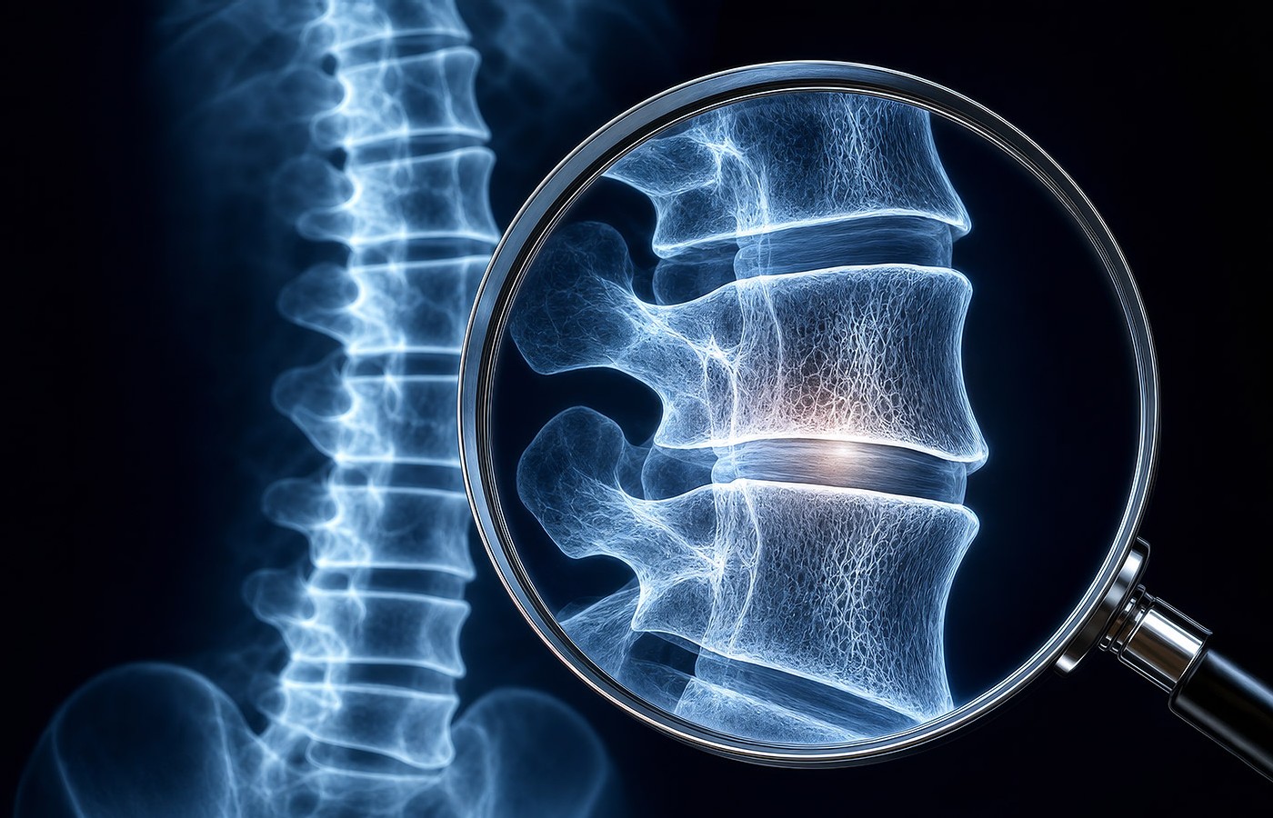
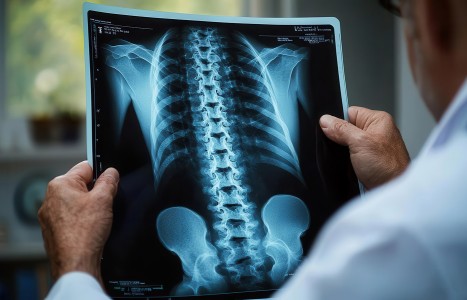
One of the most dangerous assumptions in spine care is believing that clinically significant pathology is always accompanied by obvious symptoms or classic red flags. It is not.
Every chiropractor who has practiced long enough has encountered a patient whose radiographs revealed findings that were completely unexpected. Some patients present with minimal discomfort. Others arrive for routine maintenance care. Still others report only mild stiffness or intermittent pain. Yet imaging may reveal significant structural compromise capable of altering treatment decisions immediately.
Perhaps no condition illustrates this reality better than the vertebral compression fracture.
The Silent Fracture Problem
Many healthcare providers associate compression fractures with severe trauma. In reality, vertebral compression fractures commonly occur during routine activities of daily living and may develop with little or no memorable injury.
Patients frequently attribute symptoms to aging, arthritis, muscle strain, or poor posture. Some experience only mild back pain. Others have no pain at all. The problem is that the absence of symptoms does not mean the absence of structural failure.
Research has repeatedly demonstrated that vertebral compression fractures are among the most underdiagnosed conditions affecting the aging spine. Many fractures are discovered only after radiographic evaluation is performed for unrelated reasons.
This creates an important question for chiropractors: How can a doctor determine whether a vertebral body is structurally intact without imaging? The answer is simple. They cannot.
A Common Condition Hiding in Plain Sight
Compression fractures become increasingly common with age, particularly in individuals with osteopenia or osteoporosis.
In the nationally representative NHANES 2013-2014 study involving more than 3,300 U.S. adults ages 40 and older, the overall prevalence of vertebral fractures was 5.4%. Prevalence increased dramatically with age, rising to 11% among adults ages 70-79 and 18% among those ages 80 and older.1
These findings are particularly important because vertebral fractures are frequently silent. Cosman and colleagues noted that many vertebral fractures never come to clinical attention despite being associated with increased mortality, chronic pain, disability, reduced quality of life, and substantially increased risk of future fractures.1
Other published literature suggests approximately one in four women over age 50 will sustain a vertebral compression fracture during their lifetime.4
What makes these statistics especially important is that many patients never seek care because they do not recognize a fracture has occurred. They assume they are simply getting older. Yet a structurally compromised vertebra remains structurally compromised whether symptoms are present or not.
Why Symptoms Are Not Enough
A previous movement within portions of healthcare has suggested radiographs should be reserved only for patients presenting with obvious red flags. While that concept may sound reasonable on the surface, vertebral compression fractures expose one of its major weaknesses.
Many compression fractures do not present with dramatic neurological findings, severe pain, bowel or bladder dysfunction, or other traditional warning signs. Some patients report only mild discomfort. Some report intermittent symptoms. Some deny pain entirely.
If chiropractors rely exclusively upon symptom presentation, a substantial number of compression fractures may remain unidentified. Applying spinal manipulation to an unrecognized fracture site could have significant consequences for the patient.
Radiographs provide information that history, palpation, orthopedic testing, and movement assessment simply cannot provide.
This becomes increasingly important after age 50, when the prevalence of osteoporosis, osteopenia, degenerative instability, and occult vertebral fracture rises substantially.
Perhaps the most surprising finding from the NHANES study was that most vertebral fractures occurred in patients who did not meet densitometric criteria for osteoporosis. Among all subjects with vertebral fractures, only 26% demonstrated osteoporosis by bone mineral density testing. Even among adults ages 65 and older with vertebral fractures, only 38% met osteoporosis criteria.3
In other words, the majority of vertebral compression fractures occurred in patients who would not have been identified as osteoporotic based upon bone density testing alone.
This reinforces an important clinical reality: Compression fracture risk extends well beyond patients with established osteoporosis.
The National Osteoporosis Foundation Position
The National Osteoporosis Foundation recognized the importance of vertebral fracture detection in its 2014 Clinician’s Guide to Prevention and Treatment of Osteoporosis.5
The guideline recommends vertebral imaging in older adults meeting specific risk criteria and further states that when bone density testing is unavailable, vertebral imaging may be considered based upon age alone.2
While dual-energy X-ray absorptiometry (DXA) remains the gold standard for measuring bone mineral density, plain-film radiographs continue to provide valuable clinical information regarding vertebral integrity, compression fractures, structural deformity, and moderate-to-advanced osteoporotic change.
For chiropractors evaluating patients over age 50, radiographs may serve as an important first-line assessment tool before force-based spinal manipulation is considered.
The Aging Spine Is Different
One of the realities of clinical practice is that the spine changes with age. By age 50 and beyond, chiropractors encounter increasing rates of osteopenia, osteoporosis, compression fractures, degenerative instability, spondylolisthesis, severe degenerative disc disease, facet arthrosis, and spinal stenosis. Many of these conditions may substantially influence treatment decisions.
The question is not whether these findings exist. The question is whether the doctor knows they exist before applying force to the spine. This is why updated spinal radiographs should be strongly considered in aging patients, particularly when previous imaging is outdated or unavailable.
This is not an argument for indiscriminate imaging. It is an argument for informed clinical decision-making.
Safety, Technique Selection and Patient Care
The value of imaging extends beyond diagnosis. Radiographs help determine whether spinal manipulation is appropriate, whether low-force techniques should be considered, whether specific segments should be avoided, and whether referral or co-management is warranted.
An osteoporotic vertebra requires different considerations than a healthy vertebra. A healed compression fracture requires different considerations than a structurally normal segment. A spine demonstrating instability requires different considerations than a stable spine.
Without imaging, these distinctions may not be apparent. This is not defensive medicine. It is responsible patient care.
The Clinical Principle Remains Simple
Chiropractic adjustment is a mechanical intervention applied directly to the musculoskeletal system. Understanding the condition of that system before applying force is both logical and responsible.
The profession should not view radiographs merely as a tool for identifying pathology. They are also a tool for understanding biomechanics, structural integrity, treatment appropriateness, and patient safety.6
The aging patient sitting in the treatment room may have osteopenia. They may have osteoporosis. They may have a compression fracture they do not know exists. More importantly, they may have a vertebral fracture despite not meeting formal diagnostic criteria for osteoporosis.
The scientific literature clearly demonstrates that many vertebral fractures occur in patients whose bone density does not fall within the osteoporotic range.1
The National Osteoporosis Foundation has recognized the importance of vertebral imaging in adults over age 50, particularly when risk factors are present and bone density testing is unavailable.2,5
For chiropractors who routinely apply mechanical force to the spine, understanding vertebral integrity before treatment is not excessive, it is responsible patient care.
If your 62-year-old (or older) mother went to a DC and received an adjustment, then resulting pain developed in the area of that adjustment, and it was discovered upon imaging after the fact that your mother had an aggravated compression fracture or complications aggravated by the adjustment to an old compression fracture, would you be pissed that the DC didn’t have a radiograph on hand or didn’t order one based on her age?
Safety first. See before you adjust. Because sometimes the most important diagnosis in the room is the one that cannot be detected without imaging.
References
- Cosman F, Krege JH, Looker AC, et al. Spine fracture prevalence in US women and men aged 40 years and older: results from NHANES 2013-2014. Osteoporos Int, 2017;28(6):1857-1866.
- Cosman F, de Beur SJ, LeBoff MS, et al. Clinician’s guide to prevention and treatment of osteoporosis. Osteoporos Int, 2014;25(10):2359-2381.
- Genant HK, Jergas M. Assessment of prevalent and incident vertebral fractures in osteoporosis research. Osteoporos Int, 2003;14(Suppl 3):S43-S55.
- Lentle BC, Brown JP, Khan A, et al. Recognizing and reporting vertebral fractures. CMAJ, 2007;177(5):575-580.
- National Osteoporosis Foundation. Clinician’s Guide to Prevention and Treatment of Osteoporosis. Washington, DC: National Osteoporosis Foundation; 2014.
- Kraus SJ. The Science of Whiplash: Injury, Instability, and Impairment - A Clinical-Legal Guide. Evidentiary Analysis and Expert Testimony Q&A. World Spine Research Press, 2026.
Editor’s Note: Dr. Kraus’ new textbook, The Science of Whiplash, contains an expanded review of hundreds of published studies addressing radiology related to whiplash and instability factors related to whiplash, chronicity, biomechanics, central sensitization, posttraumatic stress, and long-term outcomes. Visit www.drstevekraus.com to review the expanded table of contents.