With piriformis syndrome serving as a well-established model of myogenic pseudo-radiculopathy in the lower extremity [pt. 3 in this series], the absence of a comparable diagnostic framework for the upper extremity represents a significant clinical gap. Patients presenting with arm pain, paresthesia or perceived weakness are frequently diagnosed with cervical radiculopathy based on imaging findings alone, despite symptom patterns that do not conform to dermatomal distributions or demonstrate progressive neurological deficit.
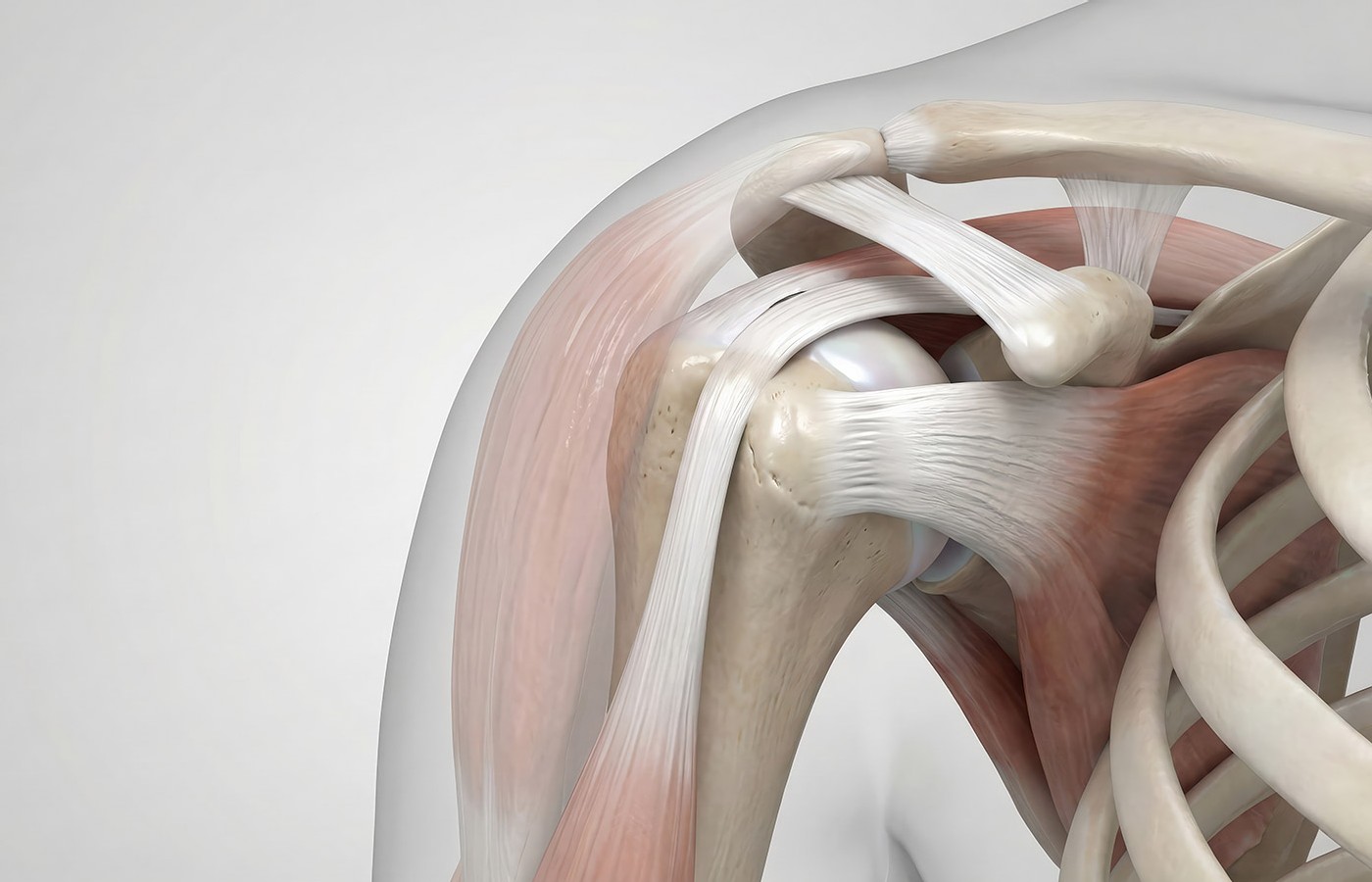
Subscapularis syndrome is introduced here as a clinically relevant and underrecognized source of upper-extremity pseudo-radiculopathy. Similar to piriformis syndrome, it reflects a functional disorder in which deep stabilizing muscle dysfunction – rather than primary nerve-root compression – drives symptoms that mimic radiculopathy.
The Role of the Subscapularis in Shoulder Stability
The subscapularis is the largest and most powerful rotator-cuff muscle, functioning as a primary anterior stabilizer of the glenohumeral joint. Its role extends beyond internal rotation to include dynamic stabilization during overhead motion, deceleration, and force transfer between the trunk and upper extremity. Unlike more superficial shoulder musculature, the subscapularis exhibits sustained tonic activity and plays a critical role in maintaining joint congruency.
This stabilizing role places the subscapularis at particular risk for overload when normal scapulothoracic and glenohumeral mechanics are disrupted. In the presence of altered scapular positioning, timing or force coupling, the subscapularis is often recruited excessively to compensate for lost stability. Over time, this compensation becomes maladaptive, resulting in hypertonicity, trigger-point development and altered neuromuscular control.
Clinical Presentation, Diagnostic Pitfalls
Patients with subscapularis syndrome rarely present with isolated shoulder pain. Instead, they often describe diffuse upper-extremity symptoms, including vague arm pain, deep anterior or posterior shoulder discomfort, fatigue with use, or intermittent paresthesia extending distally into the forearm or hand. These symptoms may fluctuate with activity level, repetitive upper-extremity use or sustained postures rather than isolated cervical spine movement.
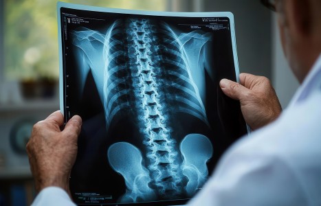
Despite this, cervical imaging frequently reveals degenerative findings that appear to offer a convenient explanation for the patient’s complaints. Disc bulges, foraminal narrowing and osteophytic changes are common in asymptomatic individuals, yet they are often interpreted as causative when arm symptoms are present. This creates a diagnostic bias that favors spinal pathology even when symptom behavior does not align with true radiculopathy.
In subscapularis-driven pseudo-radiculopathy, objective neurological findings are typically absent. Dermatomal sensory loss is inconsistent, reflexes remain intact and myotomal weakness is non-progressive. Symptoms are more likely to be reproduced with shoulder loading, scapular positioning or resisted internal rotation than with isolated cervical provocation. These features should prompt clinicians to broaden the differential diagnosis rather than escalate spine-directed care.
Parallels With Piriformis Syndrome
The clinical parallels between piriformis syndrome and subscapularis syndrome are striking. Both involve deep stabilizing muscles with high tonic demand, both demonstrate referral patterns that extend distally and mimic radiculopathy, and both are frequently identified in the presence of coincidental spinal imaging abnormalities. In each case, the muscle serves as a primary pain generator while spinal findings function as secondary or incidental contributors.
Just as piriformis syndrome reframed the understanding of sciatic-like pain without lumbar nerve-root compromise, subscapularis syndrome provides a framework for interpreting upper-extremity symptoms that do not behave like cervical radiculopathy. Recognizing this parallel allows clinicians to apply consistent diagnostic reasoning across regions rather than defaulting to a spine-centric explanation for all limb pain.
Primary vs. Secondary Drivers of Symptoms
A key diagnostic concept within subscapularis syndrome is the distinction between primary and secondary contributors to pain. Cervical joint dysfunction, degenerative disc changes or postural abnormalities may coexist with subscapularis dysfunction, but they do not necessarily represent the dominant pain generator. When the subscapularis is functioning as a compensatory stabilizer due to altered shoulder mechanics, it may drive symptoms even in the presence of cervical pathology.
This distinction is critical for case management. Treating secondary findings while ignoring the primary driver often results in incomplete or transient improvement, reinforcing the perception that symptoms are refractory or require more aggressive intervention. Conversely, addressing the muscular driver can reduce symptom intensity, normalize afferent input and improve functional tolerance without invasive escalation.
The Implications for Conservative Management
When subscapularis syndrome is correctly identified, treatment is typically conservative and minimally invasive. Effective interventions focus on restoring tissue quality and neuromuscular control rather than suppressing symptoms at the spinal level. These strategies commonly include myofascial trigger-point therapy, myofascial release, active release techniques, joint manipulation, dry needling, proprioceptive neuromuscular and progressive global stretching, and progressive shoulder stabilization and activation exercises.
These approaches target the mechanisms responsible for pseudo-radicular symptoms and often produce meaningful improvement without the need for spinal injections, nerve ablation procedures or surgical consultation when neurological compromise is absent. This conservative-first model aligns with both clinical evidence and ethical responsibility to avoid unnecessary risk.
Moving Forward in the Series
Subscapularis syndrome represents an important addition to the clinical differential diagnosis of upper-extremity pain. However, identifying the muscle alone is insufficient. Understanding why the subscapularis becomes dysfunctional – and how scapular mechanics influence its role – is essential for durable outcomes.
The next installment in this series will examine scapular dyskinesis as the upstream driver of subscapularis syndrome, drawing on biomechanical principles and clinical observations from high-demand populations, including overhead athletes. This progression will further refine the diagnostic and therapeutic framework for managing upper extremity pseudo-radiculopathy.
Editor’s Note: For previous installments in this series (parts 1-3), click here.
Resources
- Kibler WB, McMullen J. Scapular dyskinesis and its relation to shoulder pain. J Am Acad Orthop Surg, 2003;11(2):142-151.
- Lewis JS. Rotator cuff related shoulder pain: assessment, management and uncertainties. Manual Ther, 2016;23:57-68.
- Simons DG, Travell JG, Simons LS. Myofascial Pain and Dysfunction: The Trigger Point Manual, 3rd Edition. Lippincott Williams & Wilkins, 2019.
- McClure P, et al. Shoulder function and 3-dimensional scapular kinematics in people with and without shoulder impingement syndrome. Phys Ther, 2006;86(8):1075-1090.
- Woolf CJ. Central sensitization: implications for the diagnosis and treatment of pain. Pain, 2011;152(3 Suppl):S2-S15.
- Bogduk N. On the definitions and physiology of back pain, referred pain, and radicular pain. Pain, 2009;147(1-3):17-19.