With femoroacetabular impingement and labral repair surgeries on a sharp rise, we need to step back and better understand not only intra-articular lesions of the hip, but also how to treat this condition effectively.
Understanding the Cause(s)
Hip microinstability is best defined as extraphysiologic hip motion that causes pain with or without symptoms of hip joint unsteadiness, and may be the result of bony deficiency and/or soft-tissue damage or loss.1 The increased joint play degrades the joint and overworks the regional supporting structures. This increased joint motion causes cumulative microtrauma and/or acute injury to the structures of the hip. A strategic approach in treatment is critical in the success of the recovery of the patient.
Microinstability could be a result of previous injury, intra- or extra-articular pathology of the hip, congenital hip dysplasia, degenerative changes, or simply joint laxity. The most common causes of hip microinstability in research performed by Cohen, et al.,2 were iatrogenicaly caused capsular insufficiency, cam over resection in 62% of patients and soft-tissue laxity in 31% of patients.2
In this same research, the patient’s most common complaint (78% of the time) was anterior hip pain; and with a subjective complaint of gait instability in 81% of patients.
Microinstability of the hip has multiple ramifications such as labral tears, femoroacetabular impingement, premature osteoarthritis, and soft-tissue injury.1 We see the increased incidence of hip microinstability with activities that require repetitive motions such as running, pivoting, cutting, or jumping.
It is essential that as practitioners, we effectively evaluate, diagnose and treat hip microinstability to prevent compensatory motion, intra-articular and extra-articular lesions of the hip joint.
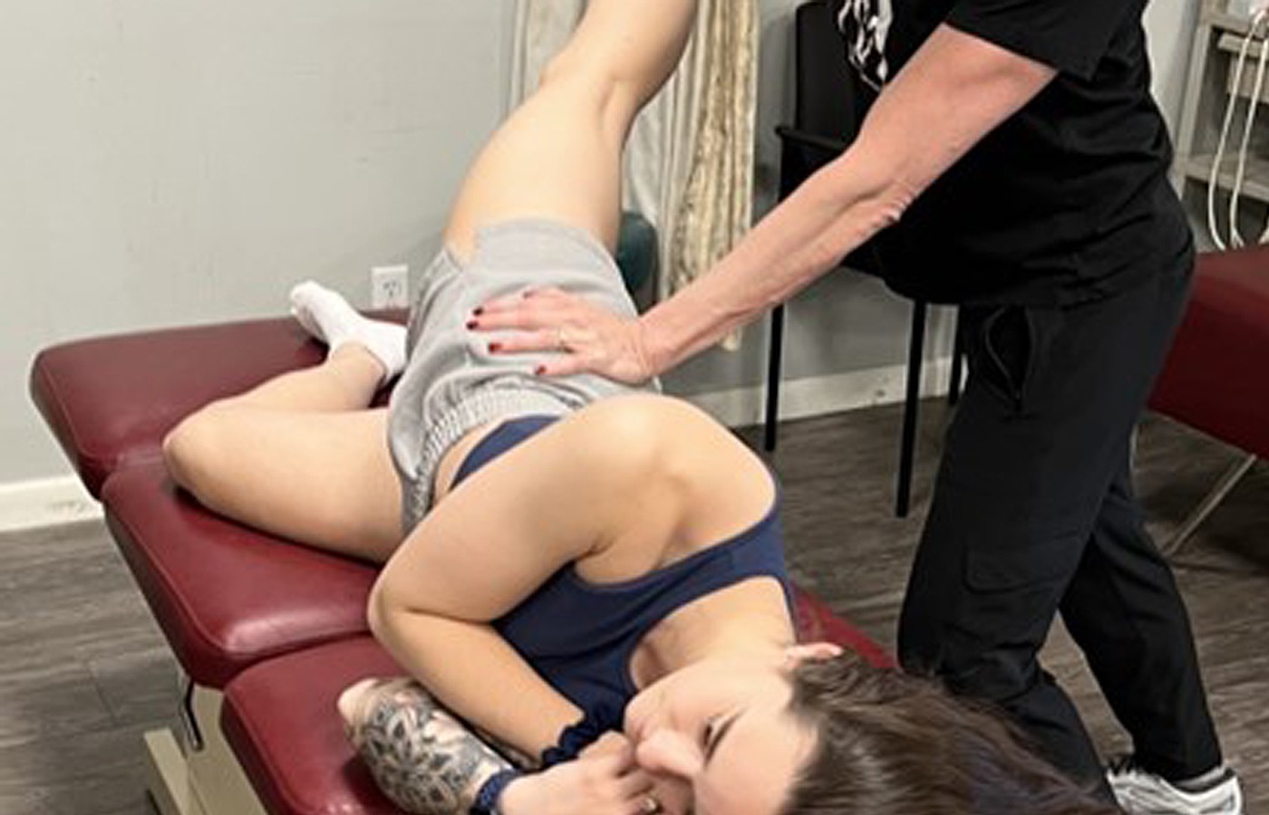
Diagnostic Considerations
Diagnosis of hip microinstability can reach a 95% confidence interval if we implement and couple three basic tests of the hip:3 the prone instability (PI) test, abduction-hip-extension-external rotation (AB-HEER) test, and hyperextension external rotation (HEER) test. Each test challenges the femoroacetabluar joint for excessive motion. A positive test is elicited when the patient reports anterior hip pain or a feeling of apprehension.
A thorough hip evaluation in addition to these hip instability tests cannot be stressed enough to further evaluate the hip for secondary or underlying conditions. This comprehensive exam should additionally include a full examination of the hip, lumbar spine, knee, ankle, foot, gait, footwear, and terrain of activity in the event of the athletic population – in other words, the entire lower limb and kinetic chain.
Additionally, digital foot scanning that visualizes all three arches of the foot to further evaluate use patterns tells a detailed story as to how the lower limb has been used and compensating for a hip condition over a period of time. This information can be used to unpeel the injury in the recovery phase of care.
Imaging of the region may demonstrate a variety of findings that could indicate the potential for microinstability of the hip. These findings can range from borderline hip dysplasia to capsular defects, capsular thinning, labral tears, and etiologies in which the femoral head is not seated deeply in the acetabulum.4
In these instances, X-ray of the region is a solid first step to evaluate the status of the hip joint and femoral head status in the acetabulum.4
MRI is a great next step to further evaluate the labrum and surrounding soft-tissue structures. Diving deeper into imaging, an MRI arthrogram can further evaluate the intra-articular status of the joint and labrum. Additionally, diagnostic intra-articular injection of anesthetic can confirm the intra-articular nature of the pathology.4
Putting together the imaging findings with the clinical findings on exam, the patient report of anterior hip pain and instability with gait, an accurate diagnosis can be reached.
One additional note here is the emerging interest in the imaging and documentation of the ligamentum teres of the hip as an underlying cause of hip microinstability. However, this procedure and findings are still emerging and have not been well-documented.5
Rehab and Recovery
Effective rehabilitation and recovery of hip microinstability consists of a multidimensional approach. Treatment can begin with palliative care and noxious activity modification; as well as the placement of a custom, flexible orthotic that supports all three arches of the foot. The idea here is to effectively support all three arches to protect the kinetic chain faults that may cause the hip to exhibit hypermobility and the patient feeling of instability with gait.
The next progression in the recovery phase is to re-establish the stability that the hip is lacking. This is executed with the initiation of stabilization training, global strengthening, and perturbation exercises. This strategic and strengthening program should focus on the dynamic stabilizers of the hip.
Keeping in mind that the hip is a multidirectional joint with muscles that work in all planes of motion, strengthening should be multidirectional, isometric and dynamic, beginning with closed-chain exercises and progressing to open-chain corrective exercises.
Finally, sport- or need-specific strengthening should be instituted early in the recovery phase in order to begin the required strength and stability gains. The inclusion of sensorimotor integration training is critical in all phases of care of the patient with microinstability of the hip.
Clinical Takeaway
The continually emerging diagnosis of hip microinstability brings with it clinical challenges for the practitioner. In patients presenting with a complaint of anterior hip pain and instability with gait, the addition of three hip stability orthopedic tests can give the practitioner confidence that this diagnosis is present.
In the corrective stage, the multidimensional approach of placing custom, flexible orthotics that support all three arches; as well as instituting corrective exercises and proprioceptive retraining for all planes of motion of the hip, can be helpful in resolving this condition.
References
- Safran MR. Microinstability of the hip - gaining acceptance. J Am Acad Orthop Surg, 2019 Jan 1;27(1):12-22.
- Cohen D, et al. Hip microinstability diagnosis and management: a systematic review. Knee Surg Sports Traumatol Arthrosc, 2023;31:16-32.
- Hoppe DJ, et al. Diagnostic accuracy of 3 physical examination tests in the assessment of hip microinstability. Orthop J Sports Med, 2017 Nov 27;5(11):2325967117740121.
- Curtis DM, et al. Hip microinstability: understanding a newly defined hip pathology in young athletes. Arthroscopy, 2022 Feb;38(2):211-13.
- Cerezal L, et al. Emerging topics on the hip: ligamentum teres and hip microinstability. Euro J Radiol, 2012; 81(12):3745-3754.