New York's highest court of appeals has held that no-fault insurers cannot deny no-fault benefits where they unilaterally determine that a provider has committed misconduct based upon alleged fraudulent conduct. The Court held that this authority belongs solely to state regulators, specifically New York's Board of Regents, which oversees professional licensing and discipline. This follows a similar recent ruling in Florida reported in this publication.
The Shoulder Abduction Test for Cervical Radicular Pathology
Described initially by Spurling as the shoulder abduction relief test,1 the shoulder abduction test is used to detect cervical radicular pathology. The test is a common part of most chiropractic curricula under another synonym (Bakody's test), and is commonly used in chiropractic practice.2-4
The shoulder abduction test is used routinely as part of orthopedic testing of the cervical spine and is specifically summoned to use if the patient reports arm pain. The patient reporting arm pain may provide proof of the test being positive during the initial history, before the test is actually employed. The patient will report the testing position as the only position in which pain is relieved and/or the only position that provides enough relief to allow sleep.
There are no contraindications to routine use of the test. It is a maneuver performed by the patient without force and is a palliative test, seeking to reduce, not reproduce symptoms. The only caution in using the test may be a patient's inability to abduct the arm on the side of testing due to another pathology or physical condition, typically of the shoulder complex.
Performing the Test

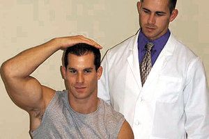
The shoulder abduction test is performed by asking the seated patient to place their hand on top of their head. This can be performed using the asymptomatic arm first to help establish a baseline finding for the side assumed to be "normal." This would be followed by alternately placing the hand of the symptomatic side on top of the head. (Figure 1) For the sake of efficiency, both hands could be placed on the top of the head simultaneously.
Other than the examination table (which should be assumed), there is no equipment required in order to perform the test. The positioning of the doctor is not a concern, as the patient would rarely need to be stabilized for the test and the doctor is not directly involved in the performance of the test. While the test is typically performed with the patient seated, it can be performed with the patient in a supine position. (Figure 2)

Once the patient is in the testing position, the position should be held for 5-10 seconds to allow symptoms to dissipate or resolve. During this time, the doctor should ask the patient, "Does this bother you or have any effect on you?" This is a good general question for orthopedic testing. The question allows the patient to respond to any symptom or change experienced. The information can then be interpreted by the doctor. The question avoids unintentionally directing the patient to a specific response, providing more objectivity.
For the test to be truly positive, the patient's arm pain must dissipate or resolve. The testing maneuver has a palliative effect on radicular arm pain. Pain should not increase if the pathology is radicular in nature, and the test is not positive if pain decreases or increases in any other areas.
A negative result does not guarantee the absence of a radicular pathology. The test should be correlated with other tests for radicular pathology and with history or other findings. Each piece of the puzzle should be contemplated individually and collectively for differential diagnosis.
Application and Confirmation
In the current environment of evidence-based health care, the utility of every procedure is being questioned. Unlike many orthopedic tests, the shoulder abduction test has been the focus of few studies. Viikari-Juntura found the test to have a specificity range of 80-100 percent and a sensitivity range of 31-43 percent.5 This can be summarized by saying the test is not seen frequently in cervical radicular conditions, but when present it is very specific in identifying the condition.
Confirming positive and negative results for the shoulder abduction test can be accomplished by additional tests for cervical radicular pathology. The two most common tests for this are the cervical compression and distraction tests. (Note: The reverse is also true: The shoulder abduction test can be utilized to confirm results for the cervical compression and distraction tests.)
The cervical compression test has a variety of synonyms and methods of performance described in the literature. For now, let's focus on the description that is most practical and functional.
The mechanism of the cervical compression test is compression of the cervical spine by the examiner. Superior-to-inferior pressure is placed on the top of the patient's head for a few seconds. This is intended to decrease the size of the intervertebral foramen, causing nerve-root irritation, and reproduces or increases the patient's arm pain.
The patient is typically seated with the head and neck in a neutral anatomical position. Variations to this test include placing the head and neck in different positions prior to applying pressure. To simplify manners, the examiner can perform the test in the neutral position first and then proceed by performing the maneuver with the head and neck flexed, extended, laterally bent and/or rotated.
Radiating arm pain is indication of a positive test. Increases or decreases in symptoms / pain in other areas are not considered to be a positive test or indicative of cervical radicular pathology.
The cervical distraction test is a palliative orthopedic test intended, like the shoulder abduction test, to relieve the symptoms of a cervical radicular pathology. The test is performed by the examiner distracting the patient's head in an inferior to superior direction. The test is meant to open or increase the diameter of the intervertebral foramen to relieve nerve-root irritation.
Differential Diagnosis and Other Considerations
Differential diagnosis is always warranted in physical testing. In this case, suspected radicular arm pain must be differentiated from thoracic outlet syndrome (TOS), myofascial referred pain and visceral referred pain.
History andPhysical Procedures Helpful in Diagnosing Cervical Radicular Pathology
|
Patients will report decreased signs and symptoms of cervical radicular pathology when sleeping with their arms above their head (a self-imposed abducted shoulder posture). Patients report increased signs and symptoms at night for TOS when sleeping with their arms above their head. In this situation, if the patient's symptoms are reproduced or intensified, the situation is deemed a reverse shoulder abduction test or reverse Bakody's test for TOS.
For efficiency in general testing, it should be noted that by having the patient place both hands on top of the head, the examiner can perform the shoulder abduction test (Bakody's) and the reverse shoulder abduction test (reverse Bakody's) simultaneously on both sides. (Figure 2)
Arm pain can also be the result of myofascial pathology, facet joint syndromes and visceral referral of pain. Palpation can help locate painful trigger points or facet joints (ligamentous / sclerotome pain) for myofascial and facet dysfunction. Charts and diagrams depicting myofascial, sclerotome and visceral referred pain are also available for differential diagnosis of these conditions.
Table 1 lists the tests and findings described to help summarize the material presented. Further reading about each of the items listed will assist the reader in the diagnosis and differential diagnosis of cervical radicular pathology.
References
- Davidson RI, et al. The shoulder abduction relief test in the diagnosis of radicular pain in cervical extradural compressive monoradiculopathies. Spine, 1981;6:441-446.
- Evans RC. Instant Access to Orthopedic Physical Assessment. St. Louis: Mosby, 2002.
- Foreman SM, Croft AC. Whiplash Injuries: The Cervical Acceleration/Deceleration Syndrome, 3rd Edition. Philadelphia: Lippincott Williams and Wilkins, 2002.
- Cipriano JJ. Photographic Manual of Regional Orthopaedic and Neurological Tests, 4th Edition. Philadelphia: Wolters Kluwer, 2010.
- Viikari-Juntura E, Porras M, Laasonen EM. Validity of clinical tests in the diagnosis of root compression in cervical disc disease, Spine, 1989;14:253-257.
- Malanga GA, Nadler SF. Musculoskeletal Physical Examination, An Evidence-Based Approach. Philadelphia: Mosby, 2006.


