It’s a new year and many chiropractors are evaluating what will enhance their respective practices, particularly as it relates to their bottom line. One of the most common questions I get is: “Do I need to be credentialed to bill insurance, and what are the best plans to join?” It’s a loaded question – but one every DC ponders. Whether you're already in-network or pondering whether to join, here's what you need to know.
Retropharyngeal Calcific Tendinitis
Retropharyngeal calcific tendinitis, also termed calcific tendinitis of the longus colli muscle, is often unrecognized or misdiagnosed because of its relatively rare occurrence. It was initially described in 1964 by Hartley. Ring, et al., determined the disorder was due to calcium hydroxyapatite deposition in the longus colli muscle.
The etiology of calcium hydroxyapatite crystal deposition is still unknown. Some suggest that repetitive trauma, recent injury, tissue necrosis or ischemia may play a role. It is believed that once the calcium crystals have deposited within the longus colli muscle, they invoke a painful, foreign body-like inflammatory response.
The condition affects adults mainly between the ages of 30 and 60. The clinical presentation includes neck pain and tenderness, limitation of motion, dysphagia, occasional mild fever, leukocytosis, and elevated erythrocyte sedimentation rate. The dysphagia is due to the close proximity of the retropharyngeal space to the adjacent pharyngeal constrictors. There may or may not be a history of trauma and hyperflexion-hyperextension injury. The typical characteristics of this entity are calcifications at the superior insertion of the longus colli tendons at the C1-C2 level and fluid collection in the retropharyngeal space.

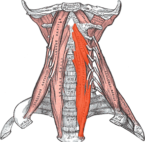
The LCM is located in the prevertebral space, extending from the anterior tubercle of the atlas to its inferior attachments at T3. It consists of the superior oblique, vertical, and inferior oblique fibers. The superior oblique fibers originate from the anterior tubercles of the transverse processes of C3-5 and insert by a tendon into the anterior tubercle on the ventral arch of the atlas. The vertical fibers arise from the bodies of C5-T3 and insert into the bodies of C2-4. The inferior oblique fibers originate from the bodies of T1-3 and insert into the anterior tubercles of the transverse processes of C5-6.
Differential diagnosis includes a retropharyngeal abscess, infectious spondylitis or neoplasm. Of course, these conditions must be ruled out before considering calcification of the LCM. The condition is self-limiting and will resolve within several weeks, but is quite painful and debilitating to the patient, especially the initial onset of symptoms. Conservative treatment, such as a short course of nonsteroidal anti-inflammatory medication (NSAIDs) and the avoidance of aggravating neck movements, can help alleviate symptoms. Knowledge of this entity is important, as it can prevent incorrect medical therapy and/or invasive treatment, such as surgical drainage of presumed present abscess.

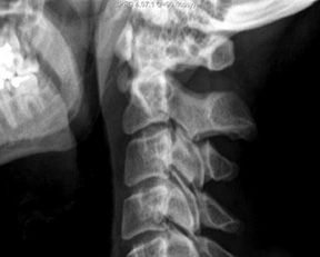
The diagnosis can be established radiographically by identification of pathognomonic findings of amorphous calcification anterior to C1-2 with associated asymmetric soft-tissue swelling. All these finding can often be identified on plain films. However, calcification may not be evident on an initial plain radiograph; only diffuse soft-tissue swelling in the prevertebral C1-4 area may provide a clue for diagnosis. A CT scan can help identify calcific deposits, which can often appear faint on plain film. MRI is not typically necessary for this diagnosis.
The same principal radiographic findings of retropharyngeal calcific tendinitis hold true for plain films, CT and MRI: prevertebral soft-tissue swelling and amorphous calcification anterior to C1-C2. The diffuse, prevertebral soft-tissue thickening typically extends from C1 to C4. The soft-tissue thickening represents either discrete effusion or diffuse edema, which can be differentiated on CT or MRI. The lack of enhancement surrounding the effusion can be helpful in differentiating a reactive effusion from an abscess. Knowledge of this condition will save a patient from unnecessary treatment and undue anxiety.
References
- Hartley J. Acute cervical pain associated with retropharyngeal calcium deposit: a case report. J Bone Joint Surg (U.S.), 1964;46:1753-1754.
- Ring D, Vaccaro AR, Scuderi G, Pathria MN, Garfin SR. Acute calcific retropharyngeal tendinitis: clinical presentation and pathological characterization. J Bone Joint Surg (U.S.), 1994;76:1636–1642.
For a more in-depth discussion, please refer to the article by Drs. Michelle A. Wessely and Timothy J. Mick in the March 2011 issue of the Journal of the Academy of Chiropractic Orthopedists.


