It’s a new year and many chiropractors are evaluating what will enhance their respective practices, particularly as it relates to their bottom line. One of the most common questions I get is: “Do I need to be credentialed to bill insurance, and what are the best plans to join?” It’s a loaded question – but one every DC ponders. Whether you're already in-network or pondering whether to join, here's what you need to know.
Modic Changes on MRI Studies
In forensic or medicolegal settings, clinicians frequently are asked to opine as to the significance of various radiographic or advanced-imaging findings. The most common of these are degenerative in nature and appear in the form of reduced disc height, osteophytes, facet joint irregularities and other signs of the normal aging process known most properly as spondylosis deformans.
Intervertebral osteochondrosis, or disc degeneration, represents a pathological process affecting chiefly the nucleus pulposus and end plates.1 This condition is quite common, being found at autopsy in more than 80 percent of people by the age of 50.2 It can be seen using plain-film radiography, as well as CT or MRI. These advanced-imaging modalities allow us to follow the progression of degenerative changes over time, but often do not allow us to definitively correlate neck pain or back pain with the spinal degenerative changes imaged.
In the field of forensic medicine, it is not uncommon for opposing experts to disagree as to the significance of findings. A common finding in lumbar MRI studies is a bulging or herniated disc. While some experts contend that herniation (i.e., more than 3 mm of protrusion) is present in up to 50 percent of asymptomatic subjects, based on an early CT study, this was a misinterpretation of the authors' findings. Subsequent studies have shown that herniations in the lumbar spine can be found in about 4 percent to 28 percent of asymptomatic adults and are more common among the elderly.3,4

It is important to correlate the patient's symptoms and clinical findings in terms of side dominance and segmental level with the finding, and it also is important to consider the patient's age. Since people are more likely to have a herniation as they age, a finding in a younger person is more likely to represent a true positive. The prevalence of herniation in the cervical spines of asymptomatic patients is much lower than in the lumbar spine and has been reported at only 4 percent to 8 percent.5
De Roos, et al., and Modic, et al., have described signal changes seen on MRI of the spine.6,7 These changes can be seen in the vertebral body adjacent to the disc(s) of interest. They have subsequently become known as Modic changes and are of three types. Type I Modic changes are considered inflammatory and are the result of fissures in the end plates. They are seen best on T2 images of the MRI and represent granulation tissue or increased vascularity. (In the presence of posterior disc bulging or herniation, disc hyperintensity on the T2 image also correlates well with a radial annular tear.)
Type II changes, which are more common, are seen chiefly on the T1 images and represent more chronic inflammatory changes and fatty infiltration of bone marrow. Type III changes correlate with extensive bony sclerosis and might show signal changes on both T1 and T2 images. These findings can be helpful in forensic applications, as the following two recent cases illustrate.
Case #1

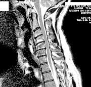
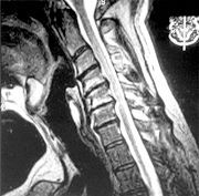
This 50-year-old male patient had a long history of neck pain and had been treated successfully in the recent past with a series of epidural steroid injections. While he was sitting in a standard office swivel chair, the base of the chair broke and he fell backward, landing on the floor. His point of contact with the floor was his upper back. His neck pain complaints and radiculopathic symptoms returned following this fall. A second course of epidural steroid injections was unsuccessful, as were all other conservative measures, and he eventually had a bi-level cervical discectomy and fusion with anterior plating. An MRI study obtained after the fall was compared to an MRI study taken a year prior to the fall and demonstrated a significant progression of the herniations at two levels and a significant progression of type I bone marrow changes in adjacent vertebral bodies (Figures 1 and 2).
The defense team argued the current complaints were similar to the complaints he'd had in the past and that, based on his previous MRI findings, this represented only a natural progression of his pre-existing condition, not a traumatic aggravation. Using measuring tools, I was able to demonstrate a three-level herniation progression of 47.5 percent, 27 percent, and 41.5 percent at the C4-5, C5-5 and C6-7 levels, respectively, over such a short period of time as would have been very unlikely absent an intervening trauma. We also were able to show the increase in the adjacent Modic changes provided additional objective evidence of a new inflammatory aggravation. At the settlement conference hearing, the defense tendered a very favorable offer.
Case #2
A 65-year-old woman was driving her vehicle at a very low speed and was struck on the passenger side by another vehicle. I served as the accident reconstructionist and biomechanist on this case. This was another case in which a long-standing spinal condition - this time lumbar - was present. Once again, the patient had been treated successfully in the past with epidural steroid injections and had a long history of recurrent low back pain. After her motor vehicle collision (MVC), she began to have more low back and leg pain. As in the first case, a second round of epidural steroid injections was unsuccessful. (Note that epidural steroid injections usually are much less effective in traumatic spinal pain versus idiopathic spinal pain, and a recent study of transforaminal epidural steroid injections has identified a shockingly high incidence of serious morbidity and mortality, suggesting a moratorium on the procedure.8)
As in case #1, Modic type I changes were evident adjacent to the offending disc and will be useful in demonstrating, objectively, a cause-and-effect relationship with her aggravation of low back and leg pain from this MVC. In people with long-standing low back pain, we are much more likely to see type II changes, which are associated with fatty marrow infiltration. The type I inflammatory changes in this case imply a recent disruption of the end-plate integrity due to the compressive, shear and bending forces imposed on the disc by the MVC. The patient subsequently underwent a fusion of L4-5. Because the case still is active, I have not included the MRI image, although the changes appear similar to those in Figures 1 and 2.
Both of these cases illustrate how Modic changes can sometimes allow us to differentiate between long-standing degenerative changes and the effects of acute trauma and subsequent inflammation. From a forensic standpoint, this differentiation can have profound implications on the outcome of a case.
References
- Ahmed M, Modic MT. Neck and low back pain: neuroimaging. Neurol Clin, 2007;25:439-71.
- Quinet RH, Hadler NM. Diagnosis and treatment of backache. Semin Arthritis Rheum, 1979;8:261-87.
- Kent DL, Haynor DR, Larson EB. Diagnosis of lumbar spinal stenosis in adults-a meta-analysis of the accuracy of CT, MR and myelography-review. Am J Roentgenol Radium Ther Nucl Med, 1992;158:1135-44.
- Boden SD, Davis DO, Dina TS, et al. Abnormal magnetic-resonance scans of the lumbar spine in asymptomatic subjects. J Bone Joint Surgery, 1990;72-A:403-8.
- D'Antoni A, Croft AC. Prevalence of herniated intervertebral discs of the cervical spine in asymptomatic subjects using MRI scans: a qualitative systematic review. J Whiplash Related Disord, 2005;5:5-13.
- De Roos A, Kressel K, Spritzer C, Dalinka M. MR imaging of marrow changes adjacent to end plates in degenerative lumbar disc disease. Am J Radiol, 1987;149:531-4.
- Modic MT, Steinberg PM, Ross JS, et al. Degenerative disc disease: Assessment of changes in vertebral body marrow with MR imaging. Radiology, 1988;166:193-9.
- Scanlon GC, Moeller-Bertram T, Romanowsky SM, Wallace MS. Cervical transforaminal epidural steroid injections: More dangerous than we think? Spine, 2007;32:1249-56.


