It’s a new year and many chiropractors are evaluating what will enhance their respective practices, particularly as it relates to their bottom line. One of the most common questions I get is: “Do I need to be credentialed to bill insurance, and what are the best plans to join?” It’s a loaded question – but one every DC ponders. Whether you're already in-network or pondering whether to join, here's what you need to know.
The Dura Mater and the Spinal Cord
This is my second article on the upper cervical spine. I'll focus here on the dura and its upper attachments. Chiropractors tend to look at the spine as 24 segments and evaluate for lack of motion between the individual segments. But trauma or poor posture can also put a stress on the dura mater, directly affecting its ability to lengthen and release. This, in turn, can affect the spinal cord, especially where the dura and cord narrows at T8-9, and where the dura attaches at the occiput, C2, the sacrum and the coccyx.
The sleeves where each nerve root exits include an extension of the dura, so any spinal level can put tension on the dura. When subluxations recur, check the dura. Treat the cause! The classic example of dural tension affecting the spine is seen in meningitis, where neuromeningeal irritation causes incredible stiffness of the neck. The dural restrictions we typically see may not be as severe as this, but have the same global effect of locking up many areas of the spine.
This concept is addressed in many chiropractic techniques, and can be called meningeal work, dural torque, or by other names. Alf Breig, a neurosurgeon, was one of the first to note how much lengthening and stretching of the spinal cord occurs in flexion, and slacking in extension. For a detailed view of the effect of trauma on the dura, I highly recommend Trauma, an Osteopathic Approach, a book by Jean-Pierre Barral and Alain Croibier (Eastland Press). Croibier does an excellent job of describing the functional anatomy of the dura and cranium, and the biomechanics of injury to the spine and cranium in trauma. The techniques we outline here are described in this text.
Supine Dural Correction
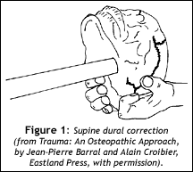
The occiput is the upper end of the spine, and the foramen magnum is the final attachment of the spinal dura. Here's a way to assess for abnormal dural tension in the upper spine. First, with the patient supine, slightly extend the head to relax the sub-occipital muscles. Second, using the soft pads of all of your fingers, pull gently superior bilaterally on the occiput. You will find tenderness at the medial occiput, unilaterally or bilaterally, when the dura is tight.
If you have not palpated for dural tension before, you will need to really tune in. It's quite a different quality of feel than palpation for joints. We are evaluating a long structure that runs from the top of the skull all the way to the coccyx. Spinal dural tension will express not as a hard articular stop, but as a rubbery, more meningeal restriction. With experience, this pull on the occiput will allow you to localize a region or level in the spine where this dural restriction is located. Fortunately, you don't have to be able to localize dural tension to a specific level to correct it. One correction is a simple cephalad pull, using ELF (engage, listen, follow) techniques to fine tune with subtle rotary or lateral bending aspects.
If you pull too hard or too fast, you will not engage the restriction you are looking for, but will go right past it, which will either have no effect, or just irritate the nervous system. Once you find the barrier back off a little to the soft edge of the barrier, and let the body release the restriction. As in all of our techniques, the key is "just enough" force. Most of us tend to pull or push too hard. By backing off to the feather edge of the barrier, we let the body participate in the correction. It's taken me years to really get this, but it's been worth it. Back off: The correction will happen easily.

Supine Dural Correction with Neck Flexion
Another way to correct dural tension is to have the patient supine, and stretch the dura by gently lifting the head into flexion. Now add lateral bending away from the side of dural tension, with rotation to further the stretch. Again, it doesn't take much force, and you need to tune in to the feel of the dura. It's different than manual stretching of the upper trapezius; you begin with flexion to the point where you feel the dural tension, then add the side bending.
Prone Correction of Dura
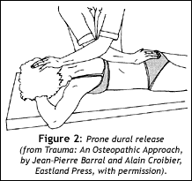
I'll mention a third dural stretch, this one done prone. Take a contact on the base of the occiput, and have your other hand directly in the midline of the spine, at T8 or T9. The picture shows a stretch to the whole of the spinal dura, from sacrum to occiput. You can cross your hands for more leverage. Here, you'll sink in to the dural level, and then traction your hands apart, lengthening the dura. Again, you may subtly add more pressure on one side or the other to add a lateral bending component. This technique uses a bit more force. You are lengthening the spine by a few centimeters, but you still want to pay attention to the barriers. You'll feel a gradual release under your hands.

Cranial Indicators
Subluxations are not limited to the spine. The dura continues beyond the occiput into the surroundings of the brain. There are many movable joints in the skull, which are well understood in the cranial manipulation world. If upper cervical problems persist, you need to check the cranium or send the patient to someone who can. Tenderness at the occipitomastoid suture (between the occiput and the temporals) strongly indicates a need for work on the cranial base.
The broader our view, and the more we know, the more patients we can help. I hope that these concepts of dural correction help you help more of your patients.
References
- Breig A. Adverse Mechanical Tensions in the Central Nervous System, John Wiley and sons, 1978
- Barral JP, Croibier A. Trauma: An Osteopathic Approach, Eastland Press, 1999.
- Heller, Marc. Low force adjusting techniques (including ELF), Dynamic Chiropractic 2001;19:26-28, http://www.chiroweb.com/archives/19/18/07.html


