Editor’s Note: The first article in this series on the pelvic floor (December 2025) focused on exercise protocols for pregnancy-related pelvic girdle pain (PPGP). Part 2 (February 2026) explored a specific combination-therapy protocol for both women and men.
Dysfunction, tension or restriction (collapsed arches, flat or rigid feet) in one area can affect the pelvic floor, contributing to symptoms such as pelvic pain, incontinence or altered pelvic posture. Note that while these connections are continuous via fascia, they involve interwoven muscles, tendons and ligaments for functional integration.
These chains allow mechanical signals, tension, and compensatory patterns in the body to ripple through the fascial system to the pelvic floor and influence function and posture.
Fascial Connections: From the Feet to the Pelvic Floor
The journey from the feet to the pelvic floor follows the lower portion of the “Deep Front Line” (DFL) described by Thomas Myers (Anatomy Trains), which provides deep support for the arches of the feet, stabilizes the legs and hips, and anchors the pelvic floor. This pathway is crucial for weight-bearing, balance, and preventing issues like flat feet or pelvic instability. Here’s the connection-by-connection breakdown:
Underside of the feet (plantar structures) to the deep posterior compartment of the lower leg: The DFL begins deep in the sole of the foot with the plantar interossei muscles and the ligamentous bed supporting the metatarsals. This connects via the tibialis posterior tendon (which supports the medial arch), and the tendons of the flexor hallucis longus and flexor digitorum longus. These structures pass behind the medial malleolus, enveloped in fascia that provides elastic recoil during walking or standing.
The fascia here links to the deep posterior compartment of the leg, between the tibia and fibula, behind the interosseous membrane, and is covered by the soleus muscle.
Deep posterior compartment of the lower leg to the back of the knee and upper leg: From the deep posterior compartment, the line ascends through the popliteus muscle and its fascia at the back of the knee, including the neurovascular bundle (tibial nerve and popliteal artery) and layers of the knee joint capsule. It then shifts medially to the adductor tubercle on the medial femoral epicondyle.
Continuing upward, the posterior track involves the adductor magnus muscle and its accompanying fascia in the posterior intermuscular septum of the thigh, running between the hamstrings and other adductors.
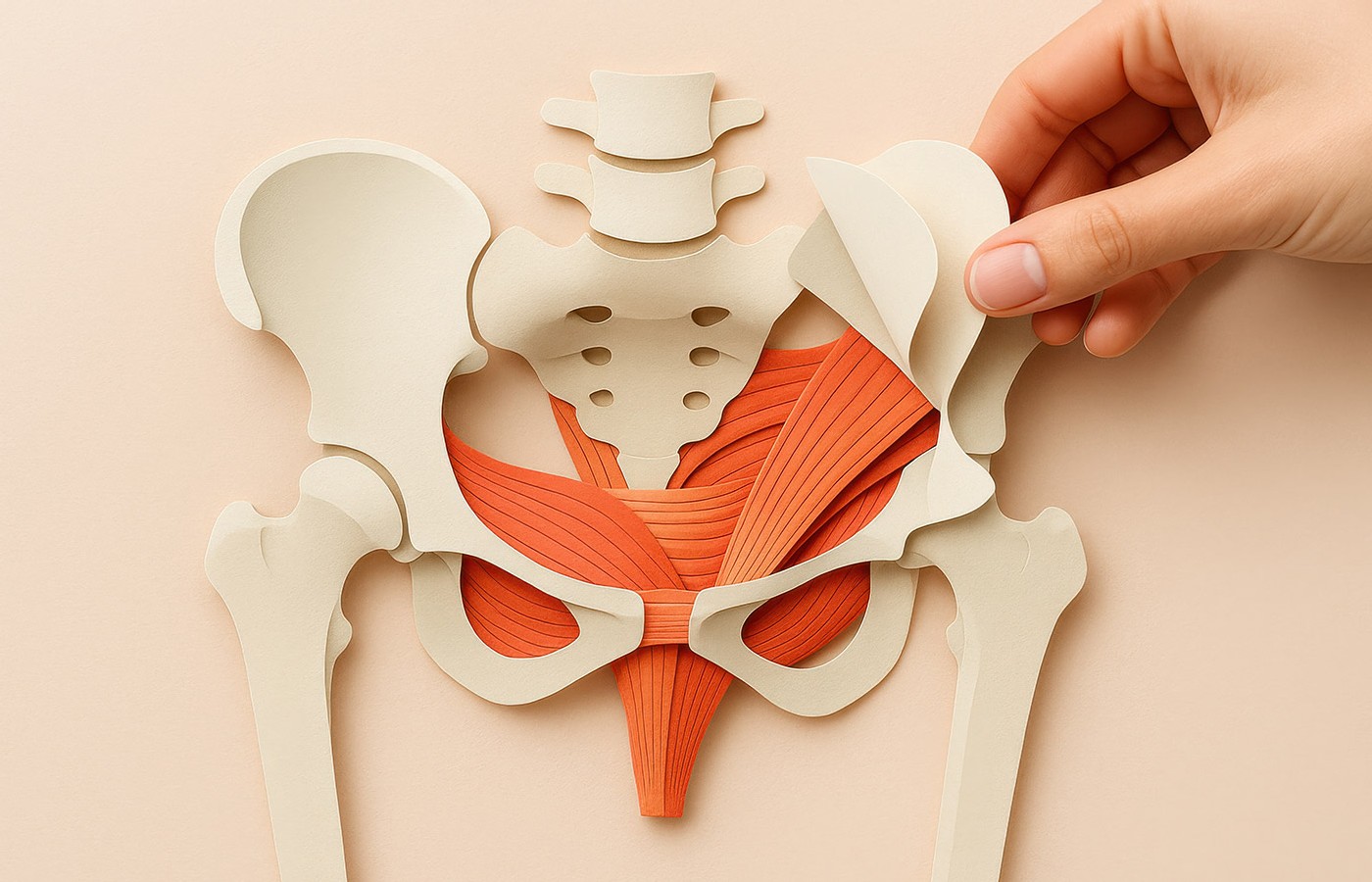
Upper thigh to the pelvis and pelvic floor: The adductor magnus and its septum connect to the medial side of the ischial tuberosity and ischial ramus. From here, strong fascial links extend over the bone to the dense covering of the obturator internus muscle (a deep hip rotator). This fascia continues via the arcuate line to the levator ani muscles of the pelvic floor, forming a stabilizing chain.
The pelvic floor itself – a muscular funnel including the coccygeus, iliococcygeus, and pubococcygeus muscles, surrounded by fascial sheets and visceral ligaments – serves as the “bottom” of the trunk’s core. It blends into the anterior longitudinal ligament on the front of the spine, providing support for the lumbar region and viscera.
This pathway highlights how foot issues (e.g., fallen arches) can pull on the fascial chain, potentially leading to knee instability, hip tightness or pelvic floor weakness.
Fascial Connections From the Tongue to the Pelvic Floor
The upper portion of the DFL links the tongue downward through the neck, thorax, and abdomen to the pelvic floor, influencing breathing, swallowing, posture, and core tension. This connection explains why tongue position (e.g., in mouth breathing or tongue ties) can affect pelvic health and vice versa. Here’s the connection-by-connection breakdown:
Tongue to the hyoid bone and suprahyoid region: The tongue connects via its intrinsic muscles (e.g., genioglossus) and the lingual septum (a fascial divider) to the hyoid bone, the small floating bone in the neck. This links to the suprahyoid muscles, including the digastric, mylohyoid, geniohyoid, stylohyoid, and even facial muscles like the masseter and buccinator. Fascial spaces in this region (e.g., pharyngeal and submandibular) provide continuity, allowing tension from the tongue to influence jaw and neck alignment.
Hyoid bone to the infrahyoid region and deep cervical fascia: Below the hyoid, the line descends through the infrahyoid (strap) muscles, such as the sternohyoid, sternothyroid, thyrohyoid, and omohyoid, which are enveloped in the deep cervical fascia. This fascia extends from the base of the skull to the upper mediastinum, connecting to the sternocleidomastoid muscle and fascial compartments like the carotid and retropharyngeal spaces.
Neurological ties, via the hypoglossal nerve (which innervates the tongue) and its links to the ansa cervicalis (cervical nerves C1-C4), further integrate this segment with lower structures.
Deep cervical fascia to the thorax and diaphragm: The deep cervical fascia continues downward into the thoracic outlet, blending with fascia around the scalene muscles, clavicle and upper ribs. It connects to the thoracic viscera and the diaphragm muscle at the base of the ribcage.
The diaphragm’s crura (tendinous pillars) attach to the lumbar spine, creating a fascial bridge that influences core pressure and posture. Innervation from the phrenic nerve (linked to cervical nerves) reinforces this, as tongue movements can subtly affect diaphragmatic function through shared neural pathways.
Diaphragm to the pelvic floor: From the diaphragm, fascial continuity extends through the psoas major muscle (a deep hip flexor attached to the lumbar spine) and into the abdominal cavity, blending with the pelvic diaphragm. This includes the levator ani and coccygeus muscles of the pelvic floor, which form a supportive sling for the pelvic organs. The connection is mediated by the anterior longitudinal ligament along the spine and visceral fascia around the abdomen, ensuring coordinated function during breathing and core activation. For instance, diaphragmatic tension (e.g., from shallow breathing) can pull on this chain, leading to pelvic floor overactivity or weakness.
This upper pathway underscores the holistic nature of the body: A restricted tongue (e.g., from a tongue tie) might contribute to neck tension, poor breathing, and pelvic floor issues like incontinence or pain.
Summary / Takeaway
I think the take-home message is how these fascial chains illustrate the body’s interconnectedness, meaning treating one end (e.g., foot exercises or tongue release) may benefit the other.
| Pathway Part |
Anatomical Details |
Functional Role |
| Plantar Fascia |
Thick sole band supporting the foot arch |
Load transfer and foot stability during gait |
| Leg Fascia (Crural and Lata) |
Deep fascial layers surrounding leg / muscles |
Force transmission, muscle compartment stability |
| Adductor Fascia |
Medial thigh fascial covering adductors |
Connects lower-limb tension to pelvis |
| Pelvic Floor Fascia |
Endopelvic fascia supporting pelvic muscles |
Pelvic organ support, tension modulation |
| Lingual / Pharyngeal Fascia |
Fascia enveloping tongue and throat muscles |
Links head and neck movement to core |
| Pretracheal Fascia |
Deep cervical fascia around trachea and thyroid |
Connects neck fascia to thoracic cavity |
| Diaphragm Center / Tendon |
Central fibrous tendon of the diaphragm |
Core fascial integration, respiratory support |
| Pelvic Fascia |
Endopelvic and perineal fascia |
Supports pelvic floor, continence, and pressure regulation |
Resources
- Myers T. Anatomy Trains: Myofascial Meridians for Manual and Movement Therapists. Churchill Livingstone, 2001.
- Primal Pictures (3-D anatomy resource): https://primalpictures.com.