It’s a new year and many chiropractors are evaluating what will enhance their respective practices, particularly as it relates to their bottom line. One of the most common questions I get is: “Do I need to be credentialed to bill insurance, and what are the best plans to join?” It’s a loaded question – but one every DC ponders. Whether you're already in-network or pondering whether to join, here's what you need to know.
Digital X-Rays
Before the early 1980s, most medical imaging utilized photographic film for reception, storage, and as a display medium for X-ray images. In fact, X-rays were discovered due to their effect on photographic emulsion. Even when ultrasound scanning was developed, the images were transitory and displayed on a monitor. Computed tomography (CT) and magnetic resonance (MR) images were originally archived on large tapes, but were stored on film, and the tapes were reused.
However, with the advent of computerized tomography and magnetic resonance, storing digital images on film has started to fade out because of lack of storage and the need to send them to various sites. There are also so many images created by CT and MR that it has become ridiculous to attempt to store all of this information on hard copy. Therefore, most of the images already in digital form are archived in their original format or compressed for storage on disc. With the current expansion in the variety of digitally acquired images and the increasing capacity of digital storage systems, the use of photographic film is on the decline.

Filmless imaging can lead to important savings of resources and a more efficient use of time and staff. The images can be easily stored, reviewed and even sent to other locations for review and consultation. What amazed me most was not the ability to store and view the images, but the fact the images themselves were more reliable. The image quality was better; each image could be manipulated to visualize areas that would not have been possible with just a film, even with a hot light and magnifying glass. This fact struck home when I was reviewing a lumbar spine series performed in a private office. The diagnostic quality of the lumbar study was fine, but there was also a digitized copy of a lumbar study performed months prior to the plain film study, so I was able to compare the two. There was a very faint shadow adjacent to the lumbar spine, which I would normally pass off as shadows from the bowel and soft tissues in the abdomen; but upon viewing the digital image of the same projection, there were several small densities in the same location on the digitized image that were most likely kidney stones - easily visualized on the digital image. It was amazing how clear these were on the digital image, and how difficult it was to see any evidence whatsoever on the plain films. I am not suggesting everyone go to digital imaging for their X-rays. It is not cost-effective for a private practitioner, but perhaps in a few years this will not be the case.
Imagine having all of your patients' X-rays on a disc, not paying to maintain a processor, and not having any chemicals to deal with - and all the X-ray series are perfect, with no retakes. You can send the images anywhere you like, to whomever you wish to review them. You can receive a report back in minutes, not days. This is not only possible, but has been so for probably 10 years in many hospitals and large radiology centers. Because of the volume of information and the budget many institutions have available, digital technology is a must. For a private practitioner, it still is a very expensive venture.
I would like to present some of the wonderful possibilities (currently available and developing) in computed radiography (CR). I am not by any means an expert in the field - in fact, it is a separate specialty in medical imaging; its professionals are called "image management specialists."
Let me attempt to describe the possibilities that open up when we are able to digitize an image. The image data can be processed to highlight regions of interest, and irrelevant information can be suppressed. Data can be combined with other pertinent patient information and made available for review to anyone in the world, provided they have the technology to view it. The files can be quickly transmitted anywhere, over any networking connection, including conventional phone lines. The information can be archived utilizing minimal space, and retrieved within seconds. The cost of this technology is decreasing (as is the case with most technology, as it becomes increasingly available). Operating costs will decrease, because X-ray film, processing equipment, chemicals and storage space will not be necessary. Properly engineered digital detectors will make it simple to perform a study, and the resulting images will be of excellent quality (no retakes due to human error). Of course, when the system goes down, there will be delays until a technician can arrive and repair the system - but we have that same problem with any computer system.
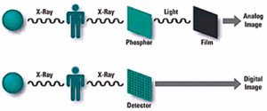
The diagram above illustrates an X-ray process resulting in an analog image; and under it, the production of a digital image.
There are many different devices available for digitizing X-ray images. Some, of course, are better than others. I would recommend waiting until the cost of these systems has decreased before considering whether you may be interested in this technology, but I guarantee you will be seeing this technology more, and it will be difficult to ignore.
For more information, check out Philip's and GE's sites (under "digital radiology") (www.gemedicalsystems.com/rad/xr/radio/products/xqi/xrd_xqi_images.html and www.medical.philips.com/main/products/xray/products/radiography/unique), both of which were gracious enough to provide me with several images I've used in recent articles. You will be amazed!
Deborah Pate, DC, DACBR
San Diego, California
patedacbr@cox.net


