It’s a new year and many chiropractors are evaluating what will enhance their respective practices, particularly as it relates to their bottom line. One of the most common questions I get is: “Do I need to be credentialed to bill insurance, and what are the best plans to join?” It’s a loaded question – but one every DC ponders. Whether you're already in-network or pondering whether to join, here's what you need to know.
Ilio-Sacral Diagnosis and Treatment, Part Three: Gluteus Medius, Piriformis and Pubic Symphysis - Positional Release and Rehabilitation Exercises
This is my final article on the ilio-sacral (IS) joint. My last article talked about IS separation, a functional diastasis in which the ilium separates laterally from the sacrum. Fully correcting IS separation requires both positional release and rehabilitation of the gluteus medius.
When the patient has IS, the joint usually is hypermobile in the basic facet direction of the SI joint, as tested from the front or back (as described in my first article in this series). The joint will test restricted in the specific lateral-to-medial direction of the IS separation, at the specific level of subluxation: S1, S2 or S3. This is a simultaneous global hypermobility of the joint, and a specific correctable restriction in the same joint.
My screening palpation for restriction is quite gentle, but when I palpate for tenderness or hypermobility, I use substantial pressure. You can make this more tolerable by pressing in slowly, rather than gouging the patient.
I cannot consistently correct IS separation strictly with an adjustment. I can't seem to eliminate or at least markedly reduce the tenderness, which is one of my cardinal signs for a successful adjustment. The tender spot for IS separation is directly over the origin of the gluteus medius muscles, particularly their posterior fibers. This is a classic muscle that gets inhibited in the late Professor Vladimir Janda's model; it also is a site for tender points in the positional release, or strain-counter-strain model.
Positional Release for the Gluteus Medius

Another technique, positional release (also known as "counterstrain") is a purely indirect technique; a "fold-and-hold" method, taking the area in the direction of ease or slack. Although positional release was invented as a structural technique, physiologically it can be seen as a way of resetting proprioceptors, primarily at tendon-osseous junctions. I tend to use it for exquisitely tender points that do not resolve with my other adjusting or fascial release techniques. An excellent organized text for this work is by D'Ambrogio and Roth.1 Positional release is one of the few manual techniques that can actually be learned from a book.
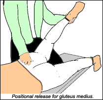
The tender points at the origin of the gluteus medius posterior are labeled as SSI, or superior sacro-iliac. Positional release is quite straightforward; you shorten the muscle or tendon until you find a position that eliminates the palpatory tenderness. It takes two contacts: one hand to lift the limb, the other to monitor the tender point. For the gluteus medius point, the patient is prone. You lift the leg into extension, abduct it, then rest it on your bent upper thigh, as shown below:
I fine-tune with medial motion of the lower leg, externally rotating the hip. Note that this combination of motions puts slack into the gluteus medius. Monitor the tender point, find the position at which it softens, and the patient's tenderness diminishes by at least two-thirds. Classic counterstrain requires you to hold this position for 90 seconds. (Yes, 90 seconds seems like forever, and some say you can shorten the time to 20 to 30 seconds by doing a brief, gentle contract-relax of the muscle involved or its antagonist.) When you are done, slowly let the leg back down. Don't let the patient help you; come out of the position with the patient as passive as possible. Retest with pressure on the tender point. When you are done, if the technique was correct, there will be a dramatic diminishing of the tenderness.
Case study: a 50-year-old woman with sudden-onset low back pain, with no recent trauma. She suffered an injury to her low back many years ago, which resolved. The only major findings on palpation were left IS separation at S2; laxity in the general motion of the left IS joint; and tenderness at the gluteus medius origin. Note that the IS separation and the gluteus medius tender points are in the same location, are easy to confuse with each other, and often exist simultaneously. In this and most cases, it took correcting both IS separation and performing counterstrain to eliminate the tenderness. To my surprise, the laxity of the SI was immediately markedly diminished.
I suspect in this case that the ligament laxity is a finding from an old injury, and was asymptomatic, well-adapted to until the recent onset of pain from some trivial, unremembered event. I suspect that the gluteal tenderness - the counterstrain points - are the glutei's inefficient efforts to substitute for the lax SI ligaments. Muscles often tighten up severely around any unstable joint. The gluteal tightness is dysfunctional, as it holds or pulls the joint apart, by pulling the ilium in a lateral direction. If I first release the muscle via positional release, my low-force adjustment of the I-S separation becomes more effective.
Rehabilitation - The Gluteus Medius and Piriformis
There are many significant muscles that affect the pelvis. For a sacro-iliac lesion, I focus on the piriformis and the gluteus medius. For any low back problem, I also address core stability provided by the transverse abdominals, multifidi and pelvic floor muscles.
You need to retrain/strengthen the gluteus medius, as it is almost always functionally weak. You can evaluate this either with Janda-style exams, as outlined in Dr. Craig Liebenson's text,2 or with classic muscle testing. The medius does not cross the SI, but it does provide frontal stability for the whole pelvis.
The piriformis is the only muscle that crosses the sacroiliac joint. It is frequently tight and short, and can directly affect the sciatic nerve. Liebenson's book outlines tests for this. If the patient feels tightness in the buttock during the piriformis stretch, it means he or she needs the stretch. There are many effective stretches for this area. I'll describe just one, my favorite, which keeps the spine in a neutral position, rather than moving it into flexion. Ask the patient to perform the following exercises:

Strengthening the Gluteus Medius
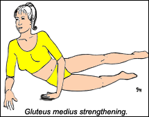
Side-lying, propped-up leg lift: Lie on your side with your upper body raised up, supported on your forearm, with your lower leg bent. Keep your hips perpendicular to the floor, and keep your upper leg in line with your body. Brace your trunk by finding neutral lumbar spine, and pulling your belly button in.
Lift your upper leg slowly straight up about eight inches, then slowly lower it. Elongate; reach out through your heel as you lift your leg. Do not twist. Rotate your leg to keep your foot parallel to the ground. You can put your upper hand on the hollow in the side of your buttock, and feel if this muscle is working as you lift. This exercise may be difficult at first. You can do this with your back and upper leg against a wall, to help keep you upright. Focus on quality, not quantity.
Lift slowly up and down, repeat 3-20 times, both sides, 1-2 times per day.
Keys - Abdominal bracing, keeping the upper leg aligned; feel the muscle work on the side of the buttock.

Pull the leg, as shown in the image above, toward the opposite shoulder.Youshould start to feel a stretch deep in the buttock muscles. At the same time,push out with your buttock muscles by pushing your tailbone toward the floorand outward. This should make the stretch stronger. You are both pulling yourleg up with your arm, and pushing your leg down toward the table, simultaneously,for the best stretch.
Hold for 30-90 seconds. Repeat 2-3 times, up to several times per day.
The Pubic Symphysis
A tender pubic symphysis is a good indicator of pubic subluxation. I palpate this from the sides of the pubic tubercle. I always use caution for this sensitive area so near the genitals: I tell my patients what I am doing; I ask permission to work on this area; and I approach with my hands from above rather than below. With males, you may want to ask them to move their "waterworks" out of the way.
The pubic tubercles, like the IS joint, have a tendency to separate, to be pulled apart, and resist medial motion. Our basic correction is to push them back together with a thumb or hypothenar contact on either side. The tubercles often also have sagittal rotation that follows the basic PI and AS pattern, and often go superior on the left. The correction that usually works is to place your hypothenar eminence on either side of the symphysis and press medially. The second corrective component is to note the direction of the superior-inferior restriction. Gently push the superior side of the tubercles inferior, and push the inferior side in a superior direction. You'll correct the pubic separation, and address the superior-inferior component.
| Subluxation | Qualities | Keys |
| shears upslip/downslip | all landmarks are inferior or superior; stiff side resists inferior or superior motion | assess both supine and prone nonphysiological shear |
| flares internal/external | ASIS stuck medial or lateral | tender medial side of ASIS |
| sagittal rotation PI and AS | usually a compensation; ASIS inferior or superior | don't correct over and over |
| ilio-sacral separation | ilium resists medial motion; can coexist with hypermobility of same side | check gluteus medius |
| pubic symphysis | separation and inferior/superior | tender lateral border of tubercles |
These three articles have attempted to provide a new view of the sacroiliac articulation, assessing the joint in multiple dimensions. If the patient does not stabilize quickly, you will need to consider other factors. You may need to add a sacroiliac belt, consider foot orthotics, and/or take a broader look at the whole spine and the lower extremities. Mineral status can be important for chronic tight muscles. Activities of daily living can undo our best adjustive techniques; especially patients who sit too long or have to repetitively bend, twist and lift. I hope this information gives you new ideas, and helps you help more of your patients. I'll finish with a table outlining the key subluxation patterns (see above).
References
- D'Ambrogio and Roth. Positional Release Therapy: Assessment & Treatment of Musculoskeletal Dysfunction, Mosby, 1996
- Rehabilitation of the Spine, A Practitioners Manual, Craig Liebenson (ed), Williams and Wilkins, 1996.
- Chouffour, Paul, Mechanical Link, North Atlantic Press, 2002
- Rex, L., Introduction of muscle energy techniques, (course notes), Edmonds, Wa., Ursa Foundation; 1996. p. 244-249
- Greenman, Philip, Principles of Manual Medicine, 2nd edition, Williams and Wilkins, 1996


