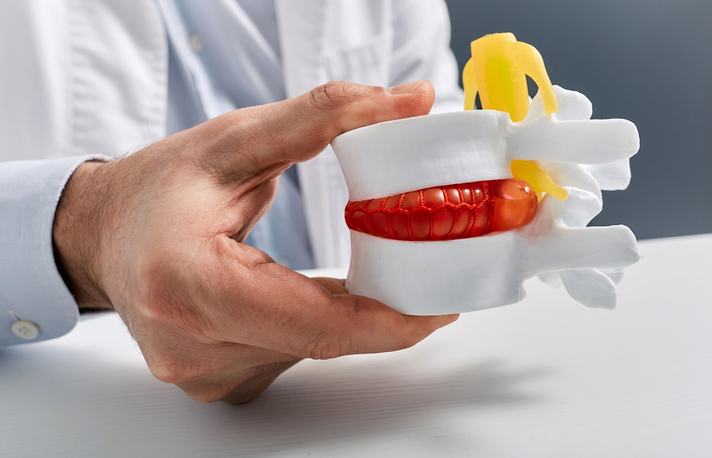
Traumatic disc herniations occur in motor-vehicle collisions (MVC). Acute traumatic disc herniations can present on MRI as protrusion, herniation, herniation with extrusion, and herniation with sequestration. A herniation can occur centrally, paracentrally, foraminally and extraforaminally.1
Disc herniations and specifically central disc herniations can be very severe and even “life threatening.” The discovery of any disc herniation should be made as quickly as possible to ensure the best patient outcome.2 Intense radicular symptoms associated with MRI-confirmed evidence of extrusion or sequestered disc fragments are accepted, straightforward symptoms of disc herniation.3 This article describes a more difficult disc herniation to diagnose, the central disc herniation (CDH).
Signs and Symptoms
A central disc herniation is associated with an elevation of the posterior longitudinal ligament (PLL).This elevation is synonymous with sprain of the PLL. The PLL sprain is indicative of an instability injury of the posterior spinal column.3 The CDH, as the diagnosis implies, involves tissue injury at the center of the posterior disc annulus.3 A CDH, due to its proximity to the spinal cord, has the potential to cause a spinal-cord injury (SCI).4
Central disc herniation can produce spinal pain and spasm typical of strain / sprain injury.5-6 A CDH also may initially cause radicular symptoms like a herniation with extrusion or one with a sequestered disc fragment.2,5
A CDH may not be large enough to directly compress the dorsal root ganglion (DRG); however, it can still produce fleeting, low-level extremity paresthesias in the first several days of recovery. Inflammatory metabolites associated with a posterior longitudinal ligament (PLL) sprain and annular injury may sensitize the DRG, giving an impression of true compression; but symptoms wane as the inflammatory stage of recovery subsides.7-8
A CDH can be linked to spinal-cord injury , anterior cord syndrome (ASAS) and cervical central cord syndrome (CCS).9-11 Headache may be present. Central cord syndrome and ASAS can manifest as extremity paresis, usually involving the upper extremities more often than the lower extremities.9-10
Anterior cord syndrome may exhibit a loss of pain and temperature sensation.9-10 Adverse bowel and bladder changes may be reported upon questioning.12 Neurogenic bowel dysfunction, dysuria and paresis are not consistent with strain / sprain injury alone.6,12
Guiliano, Guiliano and Pinto, in a unique study, utilized flexion-extension MRI to compare 100 MVC patients to 100 non-injured individuals. The study describes a 28% occurrence of acute traumatic cervical disc herniation in the MVC group. The study went on to note that 2% of the non-injured population had MRI evidence of disc herniation. A description of the herniation type was not made available in the report.3
A similar percentage of post-traumatic acute cervical disc herniations were observed in the past 1,400 MVC cases seen clinically. Most of MVC-related disc herniations diagnosed were identified as CDH or paracentral DH without evidence of DRG compression. Most patients, but not all, described an initial low-level upper and/or lower extremity paresthesia that diminished within the first two weeks.
The CDH was always a protrusion type. Thecal sac impression was not uncommon with the CDH. Ventral cord indentation, indicating a possible SCI, was observed in only a very few cases. A significantly smaller percentage of patients overall were diagnosed with extrusion or sequestered designated cervical DH. It would appear then that paracentral DH and CDH are the more prevalent DH types associated with MVC trauma.
Speeding Up the Diagnosis
The Guiliano study was performed “12-14 weeks” following the injury.3 There are a number of findings, though, that can assist the field practitioner in identifying the CDH in a more expedient manner.
A lack of improvement in the extension range-of-motion component of the initial progress examinations can be an important finding to document and a telling sign of CDH. The CDH, due to its primary posterior direction, would likely limit the patient's willingness to extend the cervical or lumbar spine when instructed to do so “just to the point of pain” in the ROM exam portion.2
Slower or non-improving pain felt in a sclerotogenous referral pattern may be associated with a PLL sprain and CDH. For most cervical PLL sprains and DH cases, this would involve pain in the upper back and neck.13
Continued pinwheel findings of hyperesthesia or even hyperalgesia on subsequent progress examinations are important clues to the discovery of CDH.4,8 This ongoing finding is likely due to the extended inflammatory stage of a HD. Herniated discs remain symptomatic longer and overall take longer to heal than more vascularized injuries such as bruising, lacerations, strain and sprain injuries.14-15
Important Diagnostic Tests
- A follow-up X-ray / dynamic X-ray of the cervical spine taken in the third week post-injury that demonstrates a 2 mm or greater anterior subluxation provides strong evidence of a PLL sprain and CDH.16
- A flexion-distraction test (FDT) noting a thoracic and/or lumbar spinal pain level finding in the third to fourth week is indicative of a CDH or other pathology.17
- A noted grip strength loss or weakness with the toe and heel walk tests is evidence of paresis, indicating potential CDH and potential SCI.9-10
Early identification of a central disc herniation will not only expedite appropriate care and recovery, but also strengthen your support of the patient's pain and injuries.
References
- “Herniated Disc: The Difference Between Bulging and Herniated Disc.” Miami Neuroscience Center. Read Here
- Sharriak S, Yasir A. Cervical Disc Herniation. Treasure Island, FL: StatPearls Publishing, LLC, 20222.
- Guiliano V, et al. The use of flexion and extension MR in the evaluation of cervical spine trauma: initial experience in 100 trauma patients compared with 100 normal subjects. Emerg Radiol, 2002 Nov;9(5):249-53.
- Schlauderaff A, Cockroft KM. Central Cord Syndrome. American Association of Neurological Surgeons. Read Here
- Pope RE. Neck Trauma: Cervical Spine Injuries. Read Here
- Freeman MD. Cervical Sprain and Strain Differential Diagnosis. Medscape, Oct. 17, 2023. Read Here
- Croft A. “Revisiting the Neurological Examination.” Dynamic Chiropractic, May 1, 2014.
- Amaya F, et al. Periganglionic inflammation elicits a distally radiating pain hypersensitivity by promoting Cox-2 induction in the dorsal root ganglia. Pain, 2009 Mar;142(1-2):59-67.
- Pearl N, Dubensky L. Anterior Cord Syndrome. Treasure Island, FL: StatPearls, 2023.
- D'Souza R. Anterior Cord Syndrome. Physiopedia.com.
- Dai L, et al. Central cord injury complicating acute cervical disc herniation in trauma. Spine, 2000 Feb 1;25(3):331-5.
- Pogio J. Neurogenic Bowel Dysfunction. Medscape.com, Feb. 10, 2023.
- Croft A. Foreman S. Whiplash Injuries: The Cervical Acceleration / Deceleration Syndrome, 2nd Edition. Baltimore, MD: Williams and Wilkins, 1995: p.p. 337-338.
- Permar G. “Herniated Disc Recovery Time. How Long Does It Take?” CrushBackPain.com, May 6, 2020. Read Here
- Barnhart, A. “How Long Does Herniated Disc Recovery Usually Take?” TheHealthBoard.com, June 7,2020.
- Freiheit T. “Diagnosing Acute Disc Herniation With the Follow-Up X-Ray (Pt.1).” Dynamic Chiropractic, May 2022. Read Here
- Freiheit T. “The Flexion-Distraction Test: A New Way to Discover Hidden Thoracic and Lumbar MVC Trauma.” Dynamic Chiropractic, March 2023. Read Here