Heel pain, worse in the morning when arising, after sitting a long time, and often loosens up after walking a bit – sounds like plantar fasciitis, right? Maybe. Let’s look at an interesting case of heel pain and how it responded to a multimodal approach to care.
Case Basics
Sam is a 60-year-old male standing 6’1” and weighing 195 lbs, who presented with pain on the bottom of his left foot and heel that had been persistent for 4-5 weeks. It began insidiously, was worse upon awakening in the morning and loosened up during the day. He felt that his shoes “pinch” and are not fitting correctly anymore.
He walks for exercise on the treadmill 2x/week at a brisk pace, with hand weights and 5 degrees elevation. In addition, he walks 20 minutes from the train station to his office every day. For work, he wears dress loafers with a thin rubber sole. Medications include losartan for hypertension, Lipitor for cholesterol management and a prescription B-complex.
Clinical Tip: The side effects of statins include musculoskeletal manifestations including tendinopathy of the rotator cuff and hand (trigger finger). Therefore, it must always be considered a potential causative agent when patients present with MSK pain. Have patients taking statins discuss a two- to four-week trial of abstinence with their PCP to determine if their MSK pain is related to a statin side effect. Taking Co-Q10 will lessen the MSK side effects.
Exam Findings
Examination revealed no nerve-root compression or traction signs, as DTRs, sensation and motor strength tests of the upper and lower extremity were WNL. His navicular drop test measured 5 mm on the right and 7 mm on the left. There was no evidence of navicular drift.
Tenderness to palpation was found over the plantar aspect and along the lateral aspect of the calcaneus, as well as over the soleus, gastrocnemius, gluteus medius, and iliolumbar ligament on the left. Orthopedic testing was positive for low back pain with Kemp’s on the left.
Restricted joint play was noted in AP glide of the talus, calcaneal eversion, AP glide of the talonavicular joint, and AP glide of the cuboid and cuneiforms on the metatarsals. In addition, the left acetabulum had reduced joint play in internal rotation and extension, and spinal pathomechanics were present at L5-S1 and the left sacroiliac joint.
Clinical Tip: In palpating the plantar fascia, initially palpate it with the foot relaxed and then re-palpate the tissues with the toes extended, which places the plantar fascia under load. When the tenderness increases under load, it indicates the plantar fascia is the potential pain driver.
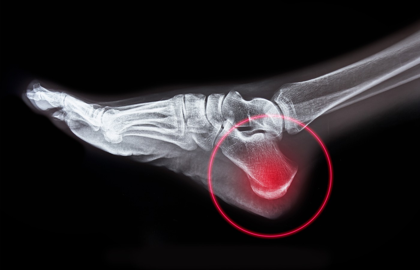
Radiographs of the left calcaneus were taken to rule out osseous pathology and demonstrated a small heel spur along the anterior-inferior aspect of the calcaneus. A comparative view of the right calcaneus was taken which indicated an identical spur; therefore, the presence of the heel spur was considered to be clinically insignificant.
Diagnosis & Initial Treatment
Plantar fasciopathy, PF, is a degenerative condition related to overuse and microtears within the plantar fascia and was the initial diagnosis. Although it may be slow to resolve, a conservative approach is the treatment of choice and fairly straightforward: improve the compliance of the plantar fascia (local modalities, stretches), release the tension within the posterior fascial subsystem (IASTM, modalities, myofascial release), balance the pathomechanics within the kinetic chain (CMT from the foot to the cervical spine as indicated), and strengthen both the intrinsic and extrinsic foot muscles.
Clinical Tip: Currently, the term plantar fasciopathy, PF, is a more accurate term versus plantar fasciitis to describe this condition.
There is good research supporting shockwave therapy for the treatment of PF – in fact, it is considered superior to cortisone injections. Using a chiropractic approach, we began Sam’s treatment with TECAR along the posterior fascial line from the hamstring to the distal metatarsals, followed by shockwave over the calf and foot.
In addition, his talus, navicular and lumbosacral regions were adjusted. Active care was stretching of the PF and calves while sitting at his desk – simply elevating the toes and dropping the heels off a 3” box under his chair. He was also advised to get shoes with a thicker sole for work.
Clinical Tip: Deep heating the soft tissues with TECAR therapy prior to shockwave enables the waves to penetrate better and according to Pratt, releases fascial densifications. Stacking the two modalities is excellent for tendinopathy, too.
Reassessment: The Missing Link
Sam’s treatment plan was weekly treatments for 6-8 weeks. After four visits, his pain had decreased in frequency, but not in intensity; and the location of pain on the plantar aspect of the heel remained the same. Considered a slow response, he was re-evaluated.
Re-examination did not demonstrate entrapment of the medial or lateral plantar nerves, radicular signs, or changes in the plantar fascia upon palpation. Loss of joint play persisted in the midfoot (navicular, cuneiform and cuboid complex) and calcaneal eversion. Therefore, CMT of the transverse tarsal (Chopat) joint was added to his treatment.
Clinical Tip: The transverse tarsal joint is formed by talonavicular and calcaneo-cuboid joints, and functions in all three planes. A transitional link between the hindfoot and forefoot, it adds to supination / pronation of the subtalar joint.
CMT of the transverse tarsal joint was the missing link in Sam’s heel pain. Once his transverse tarsal joint was adjusted, it was very cool to see the joint play in the calcaneus return to normal – immediately.
Apparently, the loss of joint play in the midfoot was affecting the rearfoot and generating excessive tension on the plantar fascia, creating his symptoms.
Clinical Tip: Wearing high-heel shoes is often prescribed for the treatment of PF. Wang determined that narrow-heel high-heel shoes actually increases the stress on the PF. Therefore, keep your patients in protective footwear and add bilateral heel lifts, as opposed to recommending wearing narrow-heel, high-heel shoes.
Stacking CMT, TECAR and shockwave is expected to restore biomechanics in a few sessions. The healing of soft tissues can take weeks to months depending on the extent of the soft-tissue compromise. Nutraceuticals, active care and modalities are excellent to enhance soft-tissue healing; however, in this case, abnormal mechanics was overloading the PF, which created the symptoms. Chiropractic CMT is indeed powerful.
Resources
- Boonchum H. Effect of a home-based stretching exercise on multisegmental foot motion and clinical outcomes in patients with plantar fasciitis. J Musculoskelet Neuronal Interact, 2020 Sep 1;20(3):411-420.
- Eliasson P, Dietrich-Zagonel F, Lundin AC, et al. Statin treatment increases the clinical risk of tendinopathy through matrix metalloproteinase release - a cohort study design combined with an experimental study, Sci Rep, 2019 Nov 29;9(1):17958.
- Lai T-W. Ultrasonography and clinical outcome comparison of extracorporeal shock wave therapy and corticosteroid injections for chronic plantar fasciitis: a randomized controlled trial. J Musculoskelet Neuronal Interact, 2018 Mar; 18(1): 47-54.
- Pratt RL. Hyaluronan and the fascial frontier. Int J Mol Sci, 2021;22:6845.
- Sun J. Extracorporeal shock wave therapy is effective in treating chronic plantar fasciitis: a meta-analysis of RCTs. Medicine, 2017;96:15.
- Wang M. The influence of heel height on strain variation of plantar fascia during high heel shoes walking - combined musculoskeletal modeling and finite element analysis. Front Bioeng Biotechnol, 2021 Dec 20;9:791238.