New York's highest court of appeals has held that no-fault insurers cannot deny no-fault benefits where they unilaterally determine that a provider has committed misconduct based upon alleged fraudulent conduct. The Court held that this authority belongs solely to state regulators, specifically New York's Board of Regents, which oversees professional licensing and discipline. This follows a similar recent ruling in Florida reported in this publication.
Atypical Femoral Fractures and Bisphosphonate Use: What to Watch For
Bisphosphonates (BP) are popular drugs, with more than 8 billion in sales in 2008; however, profits have declined as patents began expiring. Nonetheless, BP remain the most commonly prescribed drugs for patients at risk of osteoporotic fractures, with several million prescriptions written every year.
These potent inhibitors of osteoclast bone resorption definitely have demonstrated that they do reduce the risk of osteoporotic fractures, at least with short-term use. As I have discussed previously, I am not suggesting people stop taking them. However, to date, the long-term safety of bisphosphonate therapy for the treatment and prevention of osteoporosis has not been established. My main objective is to inform chiropractors of the potential problems associated with long-term use of BP, with particular attention paid to the risk of atypical femoral fracture (AFF) and the clinical signs that should elevate your suspicion.
Bisphosphonates on the FDA's Radar
In 2011, the FDA convened a joint session of the Reproductive Health Drugs and Drug Safety and Risk Management Advisory committees to examine the safety of bisphosphonates. These advisory committees examined overall bisphosphonate safety, the risk of atypical femoral fractures and osteonecrosis of the jaw, esophageal cancer risk and whether a "drug holiday" could lessen any of these risks.
There are currently four approved drugs in this class – Actonel (risedronate), Reclast (zoledronic acid), Boniva (ibandronate) and Fosamax (alendronate). While all warn of osteonecrosis of the jaw, atypical femoral fractures, esophagitis and esophageal ulcers, none carries any warning about an esophageal cancer risk. The manufacturers of these agents were uniformly opposed to label changes where there was a question as to whether or not a safety issue was clearly linked to long-term BP use. Guess we'll have to wait for this issue to be resolved sometime in the future.
Further epidemiological and clinical data are required to establish safety of BP in long-term users (>5 years). Presently, the consensus is that patients on BP should take a "drug holiday" if they have been on the drug for 3-5 years, with reassessment after they have been off the drug for 1-2 years. Those are rather broad parameters, but that is what is currently recommended, since the long-term safety of BP therapy has not been determined.
Features That Should Arouse Your Suspicion of a Fracture
Many of our older female patients are taking BP for osteoporosis. The following is a review some of the features we now know are associated with atypical femoral fracture (AFF). The following is from the American Society for Bone and Mineral Research Task Force for AFF. Findings from the first task force for AFF were published in 2010 and updated in 2013 (updated / added information in italics).
Major features: All major features should be present to satisfy definition for AFF (2010). In 2013, the task force updated some of the features of AFF changes, which are in italicized text. Note that at least four of five major features must be present. None of the minor features is required, but they have sometimes been associated with these fractures.
- Subtrochanteric location [below the lesser trochanter], or anywhere along the diaphysis [above the distal metaphysis] because these are the sites that are subject to maximal tensile loading (unlike typical hip fractures that occur at intertrochanteric level or neck of femur). The definition of AFF, the fracture must be located along the femoral diaphysis from just distal to the lesser trochanter to just proximal to the supracondylar flare.
- Minimal or no preceding trauma.
- Transverse or slightly oblique (<30°) fracture line. The fracture line originates at the lateral cortex and is substantially transverse in its orientation, although it may become oblique as it progresses medially across the femur.
- Non-comminuted or minimally comminuted.
- Complete fracture (extending through both cortices) with medial spike, or incomplete (involving only lateral cortex). Localized periosteal or endosteal thickening of the lateral cortex is present at the fracture site ("beaking" or "flaring").

- Prodromal dull or aching hip, thigh or groin pain, with history often dating back several months. Unilateral or bilateral prodromal symptoms such as dull or aching pain in the groin or thigh.
- Bilateral fractures. Bilateral incomplete or complete femoral diaphysis fractures.
- Presence of co-morbid conditions such as RA, diabetes mellitus, vitamin D deficiency, and concomitant therapy with corticosteroids, proton-pump inhibitors or other anti-resorptive agents.
- Discrete cortical thickening on the lateral side of the femur (periosteal reaction as in stress fractures) may be present. Generalized increase in cortical thickness of the femoral diaphyses.
- Delayed healing of fracture, sometimes taking several years.

Radiographic Characteristics
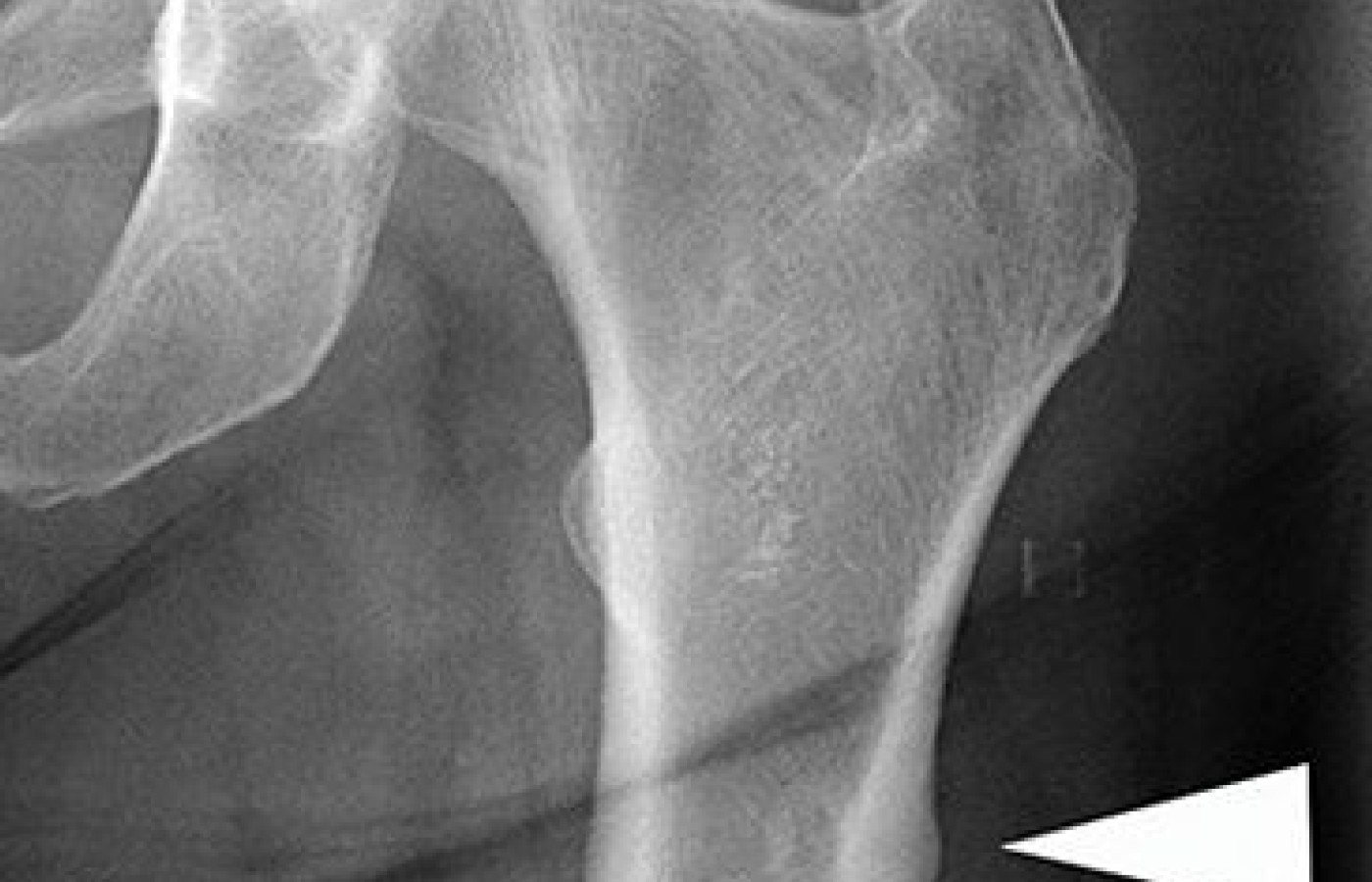
The following images are examples of what to look for if you suspect a possible stress fracture due to BP therapy. I personally would be concerned with any patient on BP therapy who complains of hip, groin or thigh pain. The prodromal period can date back several months. Even though a fracture is rare, it is better to evaluate the patient and rule out a possible fracture than assume it is simply musculoskeletal strain / sprain.
Even if you don't have your own X-ray unit, make sure you get long films of both femurs and look at the X-rays yourself. Many radiologists are still not attuned to the radiographic findings associated with AFF.
Figure 1 demonstrates the radiographic feature of lateral thickening of the cortex associated with the early development of stress fractures. Figure 2 demonstrates the radiographic findings of a horizontal stress fracture, which is a feature of these atypical fractures of the femur. Figure 3 demonstrates the classic feature of these atypical femoral fractures: a non-comminuted horizontal fracture through the femoral diaphysis.

Clinical Takeaway
The risk for atypical femoral shaft fractures increases with increased duration of bisphosphonate use; this risk decreases after cessation. As many as one in seven postmenopausal women in the United States have been treated with a bisphosphonate at some time. There is considerable controversy over the ideal duration of antiresorptive therapy, particularly since BP are not as safe as initially thought. Atypical subtrochanteric fractures, as well as osteonecrosis of the jaw, during prolonged bisphosphonate therapy have forced the FDA to re-evaluate the efficacy of continuing bisphosphonate therapy beyond three to five years.
For those patients who have discontinued bisphosphonate treatment after five years, there are currently no data to guide clinicians in determining when and whether to resume treatment. The role of repeat assessment of bone mineral density, bone turnover markers, and other clinical indicators is currently under study.
Even though chiropractors don't prescribe these drugs, we need to know what the safety issues are since many of our patients are being treated with bisphosphonates. It is my opinion that if a patient complains of groin, hip or thigh pain and they have been exposed to BP, a red flag should go up immediately. Even though a BP-induced stress fracture is rare, it should be ruled out, particularly if there is no history of trauma to the area.


