It’s a new year and many chiropractors are evaluating what will enhance their respective practices, particularly as it relates to their bottom line. One of the most common questions I get is: “Do I need to be credentialed to bill insurance, and what are the best plans to join?” It’s a loaded question – but one every DC ponders. Whether you're already in-network or pondering whether to join, here's what you need to know.
The Real Cause of Iliotibial Band Syndrome
Iliotibial band syndrome is a common injury, occurring in up to 12 percent of all runners.1 The pain associated with this syndrome is often described as "burning" and is reproduced clinically with Noble's test, in which the examiner compresses the distal band against the lateral femoral condyle while the knee is flexed 30 degrees. Although early research suggested the iliotibial band produced injury by snapping back and forth over the lateral femoral condyle (traumatizing the bursa trapped beneath), more recent research confirms that this theory is invalid.
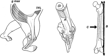
In their thorough analysis of iliotibial band anatomy and function, Fairclough, et al.,2 determine that what appears to be a forward / backward displacement of the band during knee flexion is actually an illusion created by alternating tensions generated by the tensor fasciae latae and gluteus maximus muscles. (Fig. 1) Using MRI, the authors conclusively prove the band does not snap back and forth, but is compressed into the lateral aspect of the femur as the knee is flexed, with peak compression occurring at 30 degrees flexion.

Fairclough, et al., claim the iliotibial band possesses two distinct sections: a lower ligamentous component that runs between the epicondyle and Gerdy's tubercle (which functions to limit internal tibial rotation); and a proximal tendinous component extending from the hip and attaching to the lateral femoral condyle through the dense fibrous band. (Fig. 2)

The authors claim the proximal component of the iliotibial band should be considered as a separate musculotendinous unit, suggesting the pain associated with an iliotibial band syndrome may be an enthesopathy in which tensile strain in the lower femoral attachment produces insertional bone pain (comparable to the bone pain associated with an insertional Achilles tendinitis). The authors also suggest the pain may result from repetitive compression of the highly innervated fatty tissue beneath the distal aspect of the iliotibial band. Contrary to the majority of published literature, Fairclough, et al., confirm the band itself is never inflamed, and there is no evidence of bursitis or inflammation in the distal part of the vastus lateralis muscle.
To identify biomechanical factors potentially responsible for the development of this common injury, Ferber, et al.,3 performed three-dimensional motion analysis of 35 runners with iliotibial band syndrome and compared rearfoot, knee and hip movements to 35 age-matched controls. Compared to the control group, the iliotibial band syndrome group exhibited significantly greater knee internal rotation and hip adduction, with no appreciable difference in rearfoot pronation. In fact, runners with iliotibial band syndrome had slightly reduced rearfoot eversion angles compared to the control group, which is consistent with research suggesting this injury is more likely to happen in people with high arches.
The authors state that because the band has a strong attachment to the distal femur, excessive hip adduction during stance phase increases tensile strain along the entire band, while the exaggerated knee internal rotation increases torsional strain along the distal aspect of the band. The combination of increased tensile loading from the hip and torsional loading from the knee amplifies compression of the band against the lateral knee. Because there was no difference in the angle of peak knee flexion, these authors concur with the research by Fairclough et al., questioning the validity of the sagittal plane theory in which the band snaps back and forth over the epicondyle as the knee flexes.
The detailed three-dimensional kinematic analysis performed by Ferber, et al., suggests that strengthening the hip abductors and external rotators may play an important role in the management of iliotibial band syndrome. This is consistent with research by Fredrickson, et al.,4 who demonstrated that a six-week program of hip abductor exercises produced a 51 percent increase in muscle strength, and completely resolved symptoms in 22 of 24 runners.
While the clinical efficacy of exercises may play an important role in the management of this condition, it is also essential to evaluate hip abductor flexibility, since tightness in the tensor fasciae latae, gluteus medius, and gluteus maximus muscles may increase tensile strain on the band. In an extremely detailed cadaveric study of the effect of stretching on the iliotibial band and proximal muscles, Falvey, et al.,5 surgically implanted strain gauges into the iliotibial bands of 20 fresh cadavers and evaluated the effect of three different stretches on lengthening the band. They also evaluated elasticity of the tensor fasciae latae / iliotibial band aponeurosis with ultrasonography while athletes performed maximum voluntary contractions of the hip abductor musculature.
Their detailed analysis confirmed that the iliotibial band itself is extremely rigid and resistant to stretch, since it lengthened less than 0.2 percent with a maximum voluntary contraction. Because of the extreme inflexibility of the fascial component of the iliotibial band, the authors emphasize that current treatment protocols focusing on reducing tension in the band are inappropriate, since they are treating the "symptoms of the condition rather than the cause." The authors state that to be effective, tension in the muscular component of the band must be reduced with soft-tissue therapies such as massage or dry needling of myofascial trigger points.
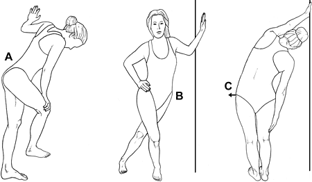
The stretches illustrated in Figure 3 are especially helpful at lengthening the upper gluteus maximus and tensor fascia latae. As demonstrated in several studies,6-8 performing deep-tissue massage prior to stretching allows for more rapid length gains. Of course, length gains can be enhanced with the use of various in-home massage tools, such as foam rollers and/or massage sticks. Although no studies have evaluated their usefulness, clinical experience confirms the various home massage tools enhance flexibility possibly by stretching the perimysium, which is considered a major extracellular contributor to passive muscle stiffness.
References
- Fredericson M, Wolf C. Iliotibial band syndrome in runners: innovations in treatment. Sports Med, 2005;35:451-459.
- Fairclough J, Hayashi K, Toumi H, et al. The functional anatomy of the iliotibial band during flexion and extension of the knee: implications for understanding iliotibial band syndrome. J Anat, 2006;208:309-316.
- Ferber R, Noehren B, Hamill J, et al. Competitive female runners with a history of iliotibial band syndrome demonstrate atypical hip and knee kinematics. J Orthop Sports Phys Ther, 2010;40:52.
- Fredericson M, Cookingham C, Chaudhari A, et al.. Hip abductor weakness in distance runners with iliotibial band syndrome. Clin J Sport Med, 2000;10:169-175.
- Falvey E, Clark R, Franklyn-Miller A, et al. Iliotibial band syndrome: an examination of the evidence behind a number of treatment options. Scand J Med Sci Sports, 2010;20:580-587.
- Hopper D, Deacon S, Das S, et al. Dynamic soft tissue mobilization increases hamstring flexibility in healthy male subjects. Br J Sports Med, 2005;39:594-598.
- Guler-Uysl F, Kozanoglu E. Comparison of the early response to two methods of rehabilitation for adhesive capsulitis. Swiss Med Wkly, 2004;134:363-368.
- Chang A, Hayes K, et al. Hip abduction moments and protection against medial tibiofemoral osteoarthritis progression. Arth Rheum, 2005;52:3515-3519.


