It’s a new year and many chiropractors are evaluating what will enhance their respective practices, particularly as it relates to their bottom line. One of the most common questions I get is: “Do I need to be credentialed to bill insurance, and what are the best plans to join?” It’s a loaded question – but one every DC ponders. Whether you're already in-network or pondering whether to join, here's what you need to know.
Centration: A Clinical Rehabilitation Approach
When rehabbing patients, there is often the question of where to start – do we stretch muscles, strengthen muscles, modify ADLs? There are many approaches and models to choose from, all of which have limitations and advantages. Joint centration integrates these models into a single functional rehab approach.
Functional joint centration, as described by Kolar, is a dynamic neuromuscular strategy that leads to the optimal joint position, allowing for the most effective mechanical advantage. Kolar illustrates that the centrated joint has the greatest interosseous contact to allow for optimal load transference across the joint and along the kinetic chain, allows for maximum muscle pull; and protects passive structures.1 Thus, decentration of a single joint affects the centration of other joints, which in turn affects the tonicity of the muscular system.2
By following this model, all movement becomes a functional test. During movement we must look at what joints become decentrated and what effects they have on the kinetic chain. Manually correct the centration with myofascial release, manipulation, MAT, etc., and evaluate the movement and kinetic chain again.
Most importantly, we must teach and exercise joints and muscles in centration. If centration during exercise is disregarded, then muscular synergy cannot be obtained. Complete body posture, not just of particular segments, is required for co-activation patterns and synergy.2 The position of the extremities or spine is just as important as the specific joints or muscles being exercised.
Applying functional joint centration to rehab protocols combines stretching and strengthening. Exercising co-activation patterns and muscular synergy restores proper muscle patterns. Stability is created in a joint, reciprocal inhibition occurs in overactive muscles, and normal function of the CNS is achieved.
When performing rehab exercises without proper centration, one is strengthening a muscle in a non-ideal position and further ingraining compensation. This compensation may decrease pain, but due to a lack of muscular synergy, faulty patterns are being trained. For example, with a shoulder dysfunction, you may be decreasing shoulder pain, but also decreasing stability of the cervical spine or function of the abdominal canister, creating a weak link in the kinetic chain.
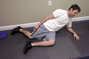
| Diagonal Sit As presented by Rich Cohen; based on concepts in movement development seen in the works of Vojta and Brunkow. This exercise activates posterior chain and abdominal function. Perform as follows: 
Notes: This should be felt in the core, glutes and lower traps. Stop if it burns more in the neck or upper traps. |
Creating centration in rehabilitative exercise is as simple as placing the patient in ideal postures to created maximum stability and power. These positions start with proper positioning of the pelvic and respiratory diaphragm, which creates stability of the lumbar spine and adequate abdominal bracing; elongation of the spine, which activates the deep stabilizing system (DNFs, multifidus, etc.); and adequate flexion, external rotation, and abduction of the shoulder and hip joints, which allows for synergy of the stabilizers and prime movers.
These postures and joint positions can be created with movement or maintained during movement. Either way, appropriate CNS programing is being trained and can be recalled during activity help keep the patient healthy and injury free.
What do these exercise postures look like? Many of these postures can be observed in the developing infant or in the partial patterns described by Dr. Vaclav Vojta. Many commonly used low-tech rehab exercises can also be modified by understanding developmental kinesiology or motor development of a child who demonstrates ideal motor patterns.1
Using the ideal movement patterns and positions described by Vojta, such as creep, roll and diagonal sit, one can use isometric contraction or movement to create properly centrated joints and synergistic muscle activation in easy-to-perform home exercises for patients.3 (See sidebar) Incorporating these principles into functional rehab programs will encourage proper CNS programming, which will influence synergistic muscle activation and motor control in our patients.
References
- Kolar P, Kobesova A. "Reflex Locomotion: Developmental Kinesiology and Stabilization Methods." Seminar series, National University of Health Sciences, 03/29/2007 - 04/01/2007.
- Liebenson C (editor). Rehabilitation of the Spine: A Practitioner's Manual. 2nd Edition. Philadelphia, PA: Lippincott Williams & Wilkins, 2007.
- Cohen R. "Introduction to Reflex Locomotion According to Vojta." Seminar series, International Vojta Society, 11/4-5/2011.


