It’s a new year and many chiropractors are evaluating what will enhance their respective practices, particularly as it relates to their bottom line. One of the most common questions I get is: “Do I need to be credentialed to bill insurance, and what are the best plans to join?” It’s a loaded question – but one every DC ponders. Whether you're already in-network or pondering whether to join, here's what you need to know.
The Upper Trapezius DOES NOT Elevate the Shoulder
How could the claim in my title be valid? Most anatomy texts state that the upper trapezius (UT) does elevate the shoulder.1-3 What about soft tissue techniques, such as postisometric relaxation or postfacilitation stretch? Should we now change our method of treating this muscle? Should we change the way we interpret the force coupling of the scapular? If you believe the contents of an excellent article, "Anatomy and Actions of the Trapezius Muscle," by Johnson and Bogduk, et al.,4 the answer is a resounding yes! The authors state that the texts do not agree with each other and "none provides reliable data on point-to-point attachments of the trapezius at a detail suitable for biomechanical modeling." They also maintain that little attention has been paid to the lines of actions of the trapezius fibers.
The authors of this study divided within human adult cadavers each fascicle (a bundle of muscle fibers with a distinct, identifiable attachment) of the trapezius, from the superior nuchal line to C7; and also the thoracic portion of the trapezius. They noted that there were no osseous origins between the occiput and the C7 spinous process. Instead, each fascicle originated from the ligamentum nuchae, not from bone. They traced each fascicle from its origin to insertion, laid down wires along the course of each fascicle, and took AP and lateral radiographs. They measured the volume and size of each fascicle to the nearest 0.5cm. All of the fascicles above the level of C7 inserted along the posterior border of the distal third of the clavicle. Fibers from C6 inserted into the distal corner of the clavicle as far as the acromioclavicular joint, while the fascicle from C7 attached to the scapula at the inner order of the acromion.

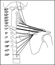
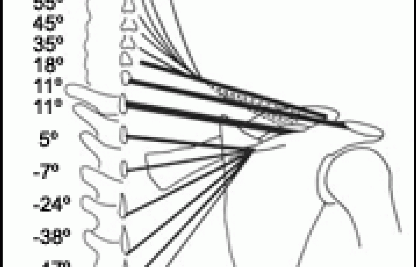
The sweep of the cervical trapezius fibers passed downward and laterally, reaching the clavicle in an almost horizontal direction. The downward orientation of the fascicles, except for those from the superior nuchal line, passed in more of a transverse than vertical direction to the clavicle. Only the fibers from the superior nuchal line showed a downward orientation that resembled an action of elevation. The illustration at left depicts the fascicles from a radiograph. The size and cross-sectional area of the fibers is also expressed by the thickness of the lines. The fascicles from the lower half of the ligamentum nuchae were much larger than the upper fibers. C6 and C7 fascicles were the largest and almost completely transverse, in orientation with the scapula in the neutral position.
It is apparent from this orientation that the nuchal portion of the trapezius is a poor elevator of the scapula, especially since "their small size limits their strength in this action"4 and the rest of the fibers have much more of a transverse direction. Also, the fibers insert on the clavicle, and not the scapula, and the action of the upper trapezius, based on its orientation, will draw the clavicle backward or medially - but not upward. These directions - backward and medial - would also be aided by the trapezius thoracic fibers, which have a transverse and upward direction.
Because the C7 and T1 fibers are close to the axis of rotation of the scapula (root of the spine of the scapula) at the beginning of shoulder abduction, their short moment arm does not participate much in early upward scapular rotation, but as the upward rotation of the scapula increases, due to the serratus anterior, the UT is able to contribute more to the force couple for upward rotation or helping in resisting downward rotation. Therefore, the idea that the trapezius by itself can act as a force couple5 (i.e., the upper fibers pulling upward, while its lower fibers pull downward) is not correct, since the upper and lower fibers of the UT do not act in opposite directions. As the serratus anterior draws the scapula laterally around the chest, the lower fibers of the trapezius act as a stabilizer and contract isometrically, while the upper fibers of the trapezius cause an upward rotation moment. So, the actions of the UT are to draw the clavicle backward or medially (rowing or pulling) and work with the serratus anterior to rotate the scapula.
Study authors Johnson, et al., account for increased EMG activity of the UT during elevation of the scapula by the UT fibers, drawing the lateral end of the clavicle medially and upward, and at the same time, causing compression load at the sternoclavicular joint. Therefore, the trapezius creates elevation by exerting an upward moment on the clavicle at the cost of compression loads at the sternoclavicular joint. The authors state that because of this mechanism, "the weight of the upper limb and any weight it carries are not borne by the cervical trapezius." The transverse UT fibers balance the movement caused by the vertical load on the shoulder, which is transferred to the sternoclavicular joint.
So, the trapezius resists lateral, instead of downward load, which is why the trapezius (C2-C6) only has to be anchored to the ligamentum nuchae, instead of the cervical spine. Since the strongest fascicles of the UT arise from C6 and C7, and the direction is almost totally transverse, the UT does not create compressive force on the cervical spine. The weight of the upper limb and the loads it carries is transferred to the sternoclavicular joint by the upper trapezius. The upper trapezius, therefore, is not an elevator of the scapula, but uses the sternoclavicular joint to sustain downward loads applied to the upper limb.
This concept of UT function may diminish its importance as a factor in the cause of cervical pain. I have noticed that the levator scapulae muscle is much more often involved than the upper trapezius, especially at its attachment to the superior medial border of the scapula.
References
- Williams PL, Warwick R. Gray's Anatomy, 36th ed., Philadelphia, WB Saunders Co. 1980:p. 566.
- Moore KL. Clinically Oriented Anatomy. Baltimore, Md., Williams & Wilkins; 1983:P.713.
- Hollinshead WH. Anatomy for Surgeons, Vol 3 - The Back and Limbs, 3rd ed. Philadelphia: Harper & Rowe, 1982:pp319-322.
- Johnson G, Bogduk N, Nowitzke A, House D. Anatomy and actions of the trapezius muscle. Clin Biomech 1994:44-50.
- Perry J. Muscle control of the shoulder. In: Rowe CR, ed. The Shoulder. New York, Churchill Livingstone; 1988:17-34.


