Some doctors thrive in a personality-based clinic and have a loyal following no matter what services or equipment they offer, but for most chiropractic offices who are trying to grow and expand, new equipment purchases help us stay relevant and continue to service our client base in the best, most up-to-date manner possible. So, regarding equipment purchasing: should you lease, get a bank loan, or pay cash?
Calcific Shadows in the Abdomen
There are numerous densities that are relatively normal findings on an AP lumbar film. Most of the time I see film that has been collimated to just about one or two inches less than actual size. This can be good and bad for several reasons: First, if the practitioner is only interested in the spine, the collimation should only include the spine and the adjacent paravertebral musculature. You'll get a better film of the osseous structures because there will be less scatter from the soft tissues of the other abdominal tissues. We know that if we take an AP lumbar on a 14 x 17, we will also get a view of the pelvis and hips; and many times that is useful for evaluating the biomechanics of the lumbar spine and pelvis. But remember this: by performing this view - AP lumbar spine and AP pelvis combined - you are basically taking a standing AP view of the abdomen. If you are taking this view you must consider all the structures demonstrated on this view, not just the osseous structures.
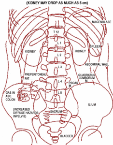
Look at the diagram of the AP abdomen view. A good way to read this is by first reviewing the soft tissue structures and organ shadows that are visible. Doing this will prevent you from just looking as the spine and missing something that may be an important clinical cause for a patient's presenting complaints. You don't have to look at this film in this manner, but it always helps to have a system that you use each time. This is my system: First, look at the lower lung fields and ribs. The lung fields should be clear and ribs should be visible and symmetrical. There shouldn't be any lucent or radiopaque densities in the ribs, and the cortex should be intact.
You may see gas in the upper margin of the stomach (magenblase), and that is normal, as long as it is below the diaphragm. Gas can be found anywhere in the colon and change the appearance of osseous structure, so remember the general path of the large bowel; and most of the time the lucent gas will not be a problem, especially if you have taken a later view.
Calcification or plaques in the great vessels can also cause some confusion on the AP, when the vessel overlies other osseous structures. Again, the lateral should resolve this confusion. The shadow of the kidneys may or may not be visible. The lower margin of the liver is often visible, but should not appear to be lower than the level of L2, otherwise it may be enlarged.
Figure 1: AP diagram of the abdomen. Depending on your technique and the patient's structural density, the shadow of many of these structures may be visible. To look at the lumbar spine, take an AP and lateral view of the lumbar region. This is best to perform on a 7X17 cassette; that way, you will not be able to over-collimate the film. Take an AP pelvis view so you can get a better view of the pelvis and hips. Many times, if the 14x17 film is not perfectly centered, the hips are excluded, and that may be the clinical cause for some of the patient's main symptoms.

|
Sometimes the shadow of the bladder can be seen, especially if it is full. Once I've reviewed the soft tissue and organ shadows, and they seem to be in their normal location, I look at the spine. I don't think I need to emphasize the osseous structures of the spine and pelvis, since we all are able to review a lumbar spine and pelvis.
I know there will be many that will disagree with this strategy, due to the different chiropractic techniques that are used to evaluate the biomechanical changes affecting the lumbar spine and pelvis. Mine is only a humble opinion that I think may help some of you with your film quality, and possibly with getting approval of films from managed care organizations. (This is another challenge altogether!)
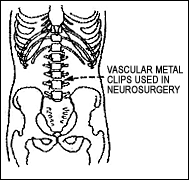
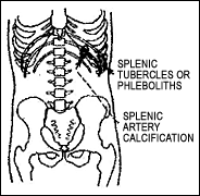
The included diagrams of common calcifications found in the abdominal region is not a complete list, but it should help you decipher most of the relatively benign findings often seen on an AP lumbar/abdomen. The only exceptions are calcifications in the pancreas, which are abnormal and are associated with pancreatic neoplasms; but fortunately, these are rather rare. Also, calcifications in suprarenal or adrenal glands may be pathologic, and should be followed, as this finding also may be associated with a neoplastic process.
When in doubt, just think about the anatomy of the abdomen and try to determine where the calcification might be anatomically. If it doesn't make sense clinically or logically, get another opinion. Who cares if it just turns out to be a normal finding? It is worth peace-of-mind! There are thousands of good general radiologists in the U.S., and most of them are pretty good at reading a simple plain film of the abdomen; as far as reviewing the biomechanics of the spine and pelvis - well, that's another issue, isn't it?

|

|

|

|

|

|


