Some doctors thrive in a personality-based clinic and have a loyal following no matter what services or equipment they offer, but for most chiropractic offices who are trying to grow and expand, new equipment purchases help us stay relevant and continue to service our client base in the best, most up-to-date manner possible. So, regarding equipment purchasing: should you lease, get a bank loan, or pay cash?
Joint Preservation Is Necessary for Hands-On Chiropractic
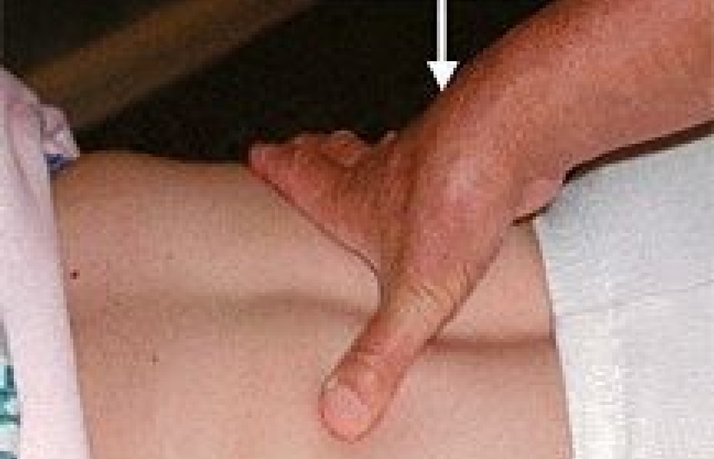
I will always remember Dr. Clarence Gonstead's hands. While his joints appeared arthritic, what was most amazing was the shape of his hands. The pisiform bone protruded; after years of adjusting, his hands had shaped into an adjusting tool. As for myself, after adjusting and performing soft tissue manipulations for the past 43 years, I suffer from a common form of osteoarthritis localized at the first carpometacarpal (CMC) joint. This joint is formed by the base of the first metacarpal and the distal side of the trapezium. There is a minimal bulge on the radial dorsal side of my CMC joint, and any stress to this joint creates pain. (See figure at below.)
Snodgrass and Rivett1 researched this problem with the CMC joint in physiotherapists and found that three recent studies revealed the hand to be the second most common anatomical site of work-related injury, following low back pain. One study reported that 91 percent of physiotherapists had to modify their treatment techniques because of thumb pain.
The thumb was not designed to be a weight-bearing joint. Many practitioners transfer their body weight through this fragile articulation up against very resistant tissue. There are significant reasons why this joint is unable to accept stress, compared to other joints of the hand or wrist. The CMC joint of the thumb is a "saddle" joint, which means each articular surface is concave in one dimension and convex in the other.
Women suffer slightly more than men with thumb problems, because the female trapezium bone is shallower, less congruent and lined with a thinner layer of articular cartilage. Functionally, the human thumb is required to be particularly mobile, and axial rotation of the thumb, as a whole, occurs primarily at the CMC joint, and in lesser amounts (accessory motions) at the metacarpal and interphalangeal (IP) joints.2
Because the articulating surfaces of this CMC joint are incongruously matched, the surface areas are not capable of continuous, solid contact when a load is transmitted through the joint. There is, therefore, increased contact stress on the small areas of the joint surfaces that do articulate.1 The CMC has a loose capsule to allow movement, and is reinforced by at least five ligaments that control the extent and direction of joint motion; help maintain normal alignment of the joint; and help control and dissipate forces produced by activated muscles.
Repetitive injury, chronic synovitis and inflammation can result in ligament laxity, causing eventual instability of the joint and leading to increased articular cartilage degeneration.3 At least 18 muscles exert force on the thumb that affects the CMC joint, and when the muscles move, there is more stress at the joint than at the distal contact point. Many soft tissue techniques require the motion of thumb flexion and adduction, and the estimated increase in compression force that occurs within the CMC joint is 12 times greater than the contact forces applied at the distal end of the thumb. "Even a low-stress act, such as brushing the teeth would, in theory, place a significant load across the joint"3 As the ligaments and muscles are stretched over time with the associated degeneration of the joint, hypermobility occurs. Increased hypermobility accelerates degenerative changes. It has been found that degenerative thumb CMC changes are occurring, even at earlier ages than expected.2

The two joints distal to the CMC, the metacarpophalangeal (MCP) and the more distal interphalangeal (IP) joint can be related to increased stress on the CMC. When a practitioner presses down directly vertical with his or her thumb, if the IP joint does not have enough extension, the MCP and CMC joints are stretched further apart, causing greater compressive loads on those joints; over time, this will lead to premature degeneration of the joint surfaces.1 Think of the manual techniques whereby the thumb is extended, putting excess stress on the MCP and CMC joints (see image above), i.e., on the belly of the subscapularis or along the paravertebral muscles. As more chiropractors realize the importance of soft tissue methods and put various techniques into practice, there will be increased chiropractic work-related thumb injuries and osteoarthritic manifestations.
As most of my readers know, I represent a producer of an instrument-assisted method of palpating and treating soft tissue lesions.4 I have personally heard from many practitioners who have used their hands for years that the technique has been a "blessing" in reducing the stress on their joints and improving their results.
References
- Snodgrass SJ, Rivett DA. Thumb pain in physiotherapists: potential risk factors and proposed prevention strategies. J of Manual & Manipulative Ther 2002; 10(4):206-217.
- West DJ, Gardner D. Occupational injuries of physiotherapists in North and Central Queensland. Aust J Physiother 2001;41(3):179-86. In: Snodgrass SJ, Rivett DA. Thumb pain in physiotherapists: potential risk factors and proposed prevention strategies. J of Manual & Manipulative Ther 2002;10(4):206-217.
- Neumann DA, Bielefeld T. The carpometacarpal joint of the thumb: stability, deformity, and therapeutic intervention. J Orth & Sports Phys Ther 2003;33(7):386-399.
- Hammer W. Graston technique: a necessary piece of the puzzle. DC Sept. 24, 2001. www.chiroweb.com/archives/19/20/03.html.


