It’s a new year and many chiropractors are evaluating what will enhance their respective practices, particularly as it relates to their bottom line. One of the most common questions I get is: “Do I need to be credentialed to bill insurance, and what are the best plans to join?” It’s a loaded question – but one every DC ponders. Whether you're already in-network or pondering whether to join, here's what you need to know.
Help for the Injured Elbow
Although the elbow and knee share certain similar anatomical characteristics, elbows in general sustain far fewer injuries. One of the main reasons for this is that non-weightbearing joints such as the elbow are usually exposed to much lower levels of force than weightbearing joints (such as the knee). There are a few sports, however, in which the elbow joint does have to support the body's weight; gymnastics is an example of such an activity.
An Integral Joint
The main duties of the elbow's joints, muscles and connective tissues are to precisely position the hand and transmit or resist a force (such as throwing a ball or spear, striking a punching bag, blocking a tackle, lifting a box, or twisting a screwdriver).1 The elbow joint is an integral part of the upper-extremity kinetic chain. Problems in the shoulder joint and cervicothoracic region can contribute to elbow joint dysfunction. Looking beyond the elbow itself for the source of its pain is critical, and any rehabilitation program designed for the elbow must address deficits in the scapular stabilizer and cervicothoracic extensor muscles, as well as proper head and shoulder posture.
Movement and Support
The humeroulnar and humeroradial articulations permit flexion and extension. The proximal radioulnar joint allows rotation (pronation and supination) to occur. Most normal activities of daily living can be performed even with partial limitation of any (or even all) of these elbow movements. However, compensations will tend to occur in adjacent body segments (such as the shoulder and spine), and performance levels in most sports will decrease quickly.2
The elbow is an inherently stable joint; however, connective tissues do provide needed additional support. These include the annular ligament (encircling the head of the radius), the medial collateral ligament (the major stabilizer against valgus stress) and the interosseous membrane (which prevents separation of the radius and ulnar shafts). If any of these connective tissues are injured, an elbow sprain is the likely result.
Most of the muscles involved in elbow function and movement originate on the humerus and insert on either the radius (biceps, brachioradialis, and pronator teres), or the ulna (brachialis, triceps, and anconeus). Two additional muscles (supinator and pronator quadratus) form a radioulnar group. Two very important elbow muscles - the wrist extensors and flexors - primarily move the hand and wrist. Manual testing can often identify in a short period of time which of these muscles are weakened and painful on contraction, indicating an elbow strain. If there is nonpainful muscle weakness around the elbow and/or wrist, a neurological condition of the lower cervical nerves (C5-8) must be considered.
How Injuries Occur
Direct trauma or overuse (due to repetitive arm and hand movements) can cause elbow injuries. Here are several common elbow injury patterns:3
- "Golfer's elbow" (medial epicondylitis) - overuse tendinosis of the wrist flexors. Stockard reports overuse injuries are "more common among amateur golfers than among professional golfers."4
- "Little League elbow" - Repetitive pitching microtrauma can cause permanent damage.
- "Nursemaid's elbow" - forced radial head dislocation in a young child (2-4 years of age).
- Olecranon bursitis - acute or repetitive direct trauma to the bursa over the olecranon.
- Panner's disease (osteochondrosis) - Overuse causes avascular damage to the capitellum.
- "Tennis elbow" (lateral epicondylitis ) - overuse tendinosis of the wrist extensors.
- Triceps tendinitis - acute or repetitive strain of the triceps insertion on the olecranon.
Elbow Sprain Rehabilitation
Damage by trauma to one or more of the connective tissues of the elbow can lead to joint instability and eventual degenerative changes. Work duties and sports activities often must be restricted to prevent further damage. Once the ligaments have undergone sufficient early repair, controlled passive motion, gentle sustained stretches, and friction massage will help to prevent the formation of adhesions. Resistance exercises are introduced to stimulate a stronger repair and assist in the remodeling process. Isometric forms progress to isotonic forms of resistance, based on the patient's tolerance for joint motion. Exercises for grip and proximal stability at the shoulder should also be included, especially for athletes.5
Elbow Strain Rehabilitation
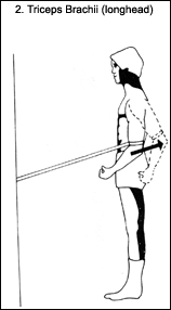
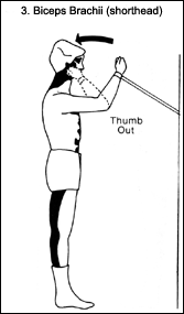
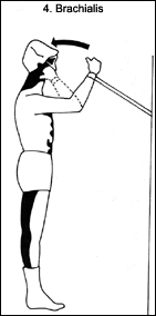
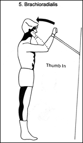
Overuse and repetitive strain are the most common sources of injury to the muscles and tendons around the elbow. For these conditions, a brief period of support and restricted activity is usually necessary.6 The use of a counterforce brace for the elbow should be implemented.7 However, controlled re-strengthening should be initiated early, with the brace on. Elastic tubing is a safe, easy method of providing progressive resistance exercises.8
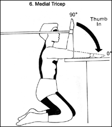
An effective elbow rehabilitation program starts with a consistent isotonic exercise routine, using elastic tubing to perform resisted pronation and supination. This should be performed initially within a limited, pain-free range of motion, building to full range as pain subsides. If the patient has tennis elbow, an overuse strain of the wrist extensors, special attention is given to these muscles. Sustained stretches are performed, followed immediately by full-range, progressive strengthening of wrist extension, with special focus on the eccentric phase of the exercise.
Eventually, the entire series of elbow exercises should be performed. This inexpensive rehabilitation program should initially be practiced under supervision to ensure proper performance. After proper exercise mechanics and control are demonstrated, a self-directed program of home exercises is appropriate.
Distortions and Alignments
Two additional factors to consider are the connection between elbow function and shoulder stability9 and the influence of cervicothoracic posture on elbow function. Specific postural distortions - such as thoracic kyphosis and cervical anterior translation (causing a "forward head") - must be addressed with corrective exercise training. An additional potential complicating postural factor is the alignment of the scapula on the thoracic cage - particularly when the shoulder is "rolled forward" (protracted). Correction of these chronic misalignments will significantly reduce the biomechanical stress during use of the arm and help prevent musculotendinous overload at the elbow.10
Repair, Then Rehab
An appropriate and progressive rehab program should be started early in the treatment of patients with elbow injuries, but only after ligaments and connective tissues have repaired sufficiently. Simple, yet effective rehab techniques are available, none of which requires expensive equipment or significant time commitments. A closely monitored home exercise program using exercise tubing is recommended, since this allows the doctor of chiropractic to provide cost-efficient, effective and specific rehabilitative care.
An important aspect of elbow rehabilitation is recognizing and addressing the biomechanical alignment problems and postural factors that can lead to substitution patterns and elbow overuse. This entails screening the patient for forward head and flexed (kyphotic) torso postures and protracted (forward) shoulders. Failure to recognize these complicating factors can result in a patient with recurring elbow complaints.

|
|
|
|
|
|

|
References
- Weisner SL. Rehabilitation of elbow injuries in sports. Phys Med Rehabil Clin North Am 1994;11:402-409.
- Nordin M, Frankel VH. Basic Biomechanics of the Musculoskeletal System, 2nd ed. Philadelphia: Lea and Febiger; 1989. 253.
- Souza TA. Differential Diagnosis for the Chiropractor. Gaithersburg, MD: Aspen Pubs; 1997. 145.
- Stockard AR. Elbow injuries in golf. J Am Osteopath Assoc 2001;101(9):509-516.
- Kibler WB, et al. Functional Rehabilitation of Sports and Musculoskeletal Injuries. Gaithersburg, MD: Aspen Pubs; 1998. 181.
- Lewis M, Hay EM, Paterson SM, Croft P. Effects of manual work on recovery from lateral epicondylitis. Scand J Work Environ Health 2002;28(2):109-116.
- Groppel JL, Nirschl RP. A mechanical and electromyographic analysis of the effects of various joint counterforce braces on the tennis player. Am J Sports Med 1986;14:195.
- Roy S, Irvin R. Sports Medicine: Prevention, Evaluation, Management, and Rehabilitation. Englewood Cliffs: Prentice-Hall;1983. 224.
- Abbott JH. Mobilization with movement applied to the elbow affects shoulder range of movement in subjects with lateral epicondylalgia. Man Ther 2001;6(3):170-177.
- Kibler WB, Press JM. Rehabilitation of the elbow. In: Functional Rehabilitation of Sports and Musculoskeletal Injuries. Gaithersburg, MD: Aspen Pubs; 1998. 171.
Kim Christensen, DC, DACRB, CCSP, CSCS
Ridgefield, Washington


