New York's highest court of appeals has held that no-fault insurers cannot deny no-fault benefits where they unilaterally determine that a provider has committed misconduct based upon alleged fraudulent conduct. The Court held that this authority belongs solely to state regulators, specifically New York's Board of Regents, which oversees professional licensing and discipline. This follows a similar recent ruling in Florida reported in this publication.
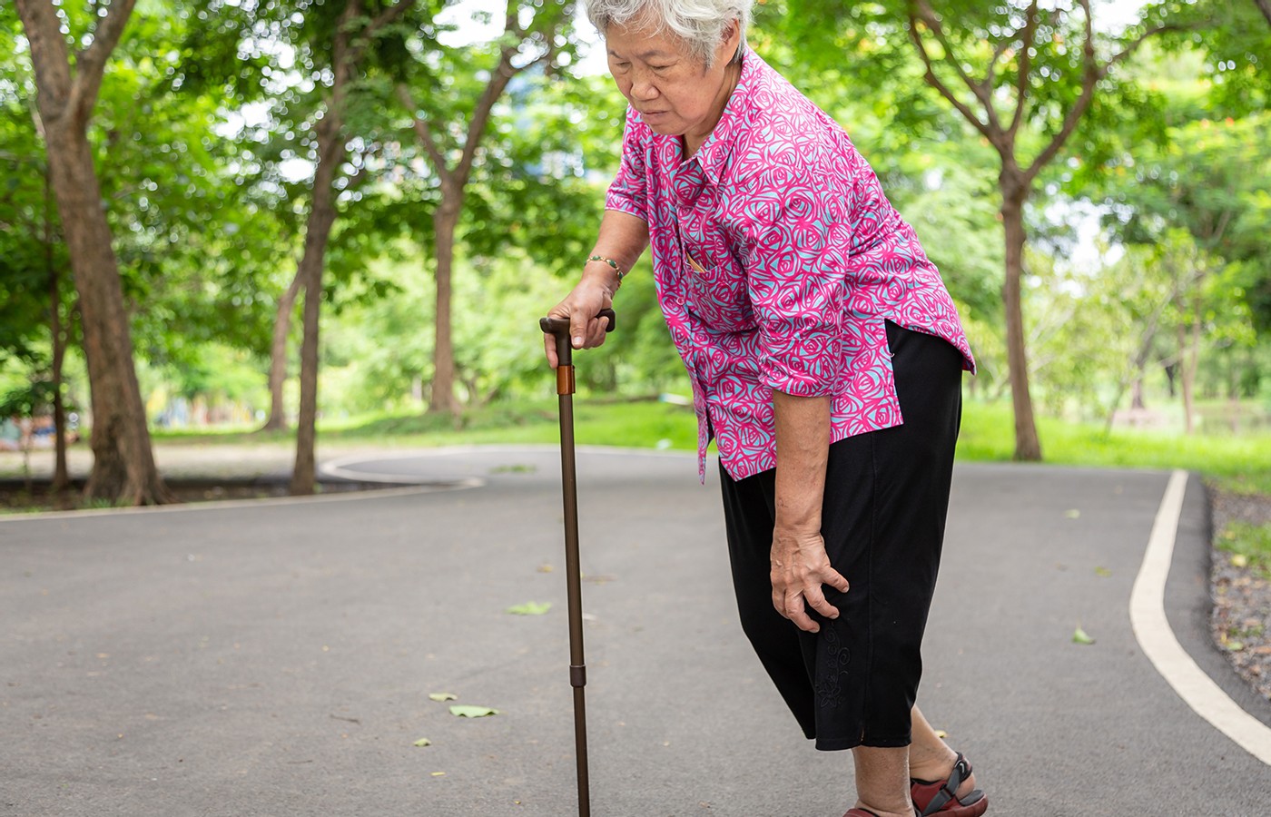
Knee Pain With Walking
- For the past two weeks, the patient has been experiencing difficulty walking because of pain in the left knee.
- The patient makes the “O” sign when describing the area of the knee pain.
- The treatment strategy included manipulation of the pelvis and left lower extremity to reduce joint dysfunction and biomechanical strain; soft-tissue treatments to reduce trigger points and pain; and locating a provider with a shockwave device.
Editor’s Note: In his last column, Dr. Lehman explored knee pain that ultimately yielded a diagnosis of synovial plica syndrome. This month, he outlines a different diagnosis for a patient presenting with knee pain. Can you figure it out?
Challenge your diagnostic skills – and perhaps rethink your differential diagnosis – of a patient who presents with a chief concern of “I have pain in my knee when I walk.” I trust this case discussion will enhance your clinical skills and benefit your patients.
History / Subjective
Chief Concern: “I have pain in my knee when I walk.”
History of Present Illness: For the past two weeks, the patient has been experiencing difficulty walking because of pain in the left knee. There is no recent history of trauma, but for the past three months he has been noticing stiffness in the left hamstrings and quadriceps muscles with his daily walking.
During his 5-7 mile walks, he would stop to stretch his hamstrings and left knee squats to reduce the discomfort, and then complete the walk. The pain is like a compression of the knee while walking. Stretching of the hamstrings and squatting reduced the pain until the past three weeks.
Unfortunately, his daily walking has ceased because of the increased pain in the left knee for the past two weeks. He points to the medial and lateral knee regions as the areas of pain.
In addition, he makes the “O” sign when describing the area of the knee pain. He denies any locking of the knee. As a teenage athlete, he did experience runner’s knee on the left.
A deep, dull ache in the left buttocks has been present with extended periods of sitting for the past two months. He has a history of statin myopathy with subsequent piriformis syndrome in the left lower extremity. The statin prescription was necessary following a coronary event with complete occlusion of the left anterior descending coronary artery. A stent implant was necessary.
Over the past six months, he gained 15 pounds and considers his weight to be too much.
Currently, sitting and walking increases the pain in the left knee, which he rates at 8/10 when attempting to walk down a decline path, or attempting to walk up or down stairs. Lying down or reclining in his lounge chair reduces the pain significantly.
There is pain and stiffness in the left knee upon rising to stand. He is unable to stand on the left foot to wash his right foot while in the shower because of pain and fear of falling.
While visiting friends in another state, he saw a chiropractor who diagnosed the condition as tendonitis and myofascial pain syndrome. The chiropractic physician applied shockwave therapy for 20 minutes. The doctor demonstrated the trigger points with the shockwave and the painful tendons.
Following the treatment, the patient was able to walk comfortably without any pain. The doctor advised him to seek additional therapy if the pain returned, which it did on the following day. But each day following the treatment, the knee pain increased.
Objective Findings
Appearance: Patient exhibits painful behavior with a limping gait (left lower extremity) upon entering the examination room.
Posture: Pelvic obliquity with a posterior-inferior left pelvis and a right anterior-superior pelvis in the standing position.
Thessaly Test: Patient is unable to perform because of severe pain in the left knee when attempting to rotate the knee internally or externally in the fully erect position. The pain is located at the medial knee, pes anserine bursa and the posterior lateral left knee.
McMurray Test: Does not exhibit any pain in the left knee.
Long Sitting Test: Supine = appearance of a short left lower extremity; seated = appearance of a long left lower extremity.
Palpation: Pain is produced with palpation of trigger points in the iliotibial band, inferior and lateral hamstrings, and the sartorius, gracilis, and semitendinosus tendons. This palpation reproduces the patient’s chief concern. Trigger points and very taut bands are also revealed in the left piriformis muscle and the iliopsoas muscle.
Palpation of the left knee joint lines does not produce pain.
Flexion, abduction, and external rotation with extension of the left hip demonstrate a reduced passive range of motion without hip pain compared to the right hip. The opposite hip is not painful with a full range of motion.
Flexion, adduction and internal rotation of the left hip demonstrate reduced passive range of motion with some discomfort in the piriformis muscle. The opposite hip is not painful with a full range of motion.
Assessment / Diagnosis
- Piriformis syndrome with resultant pelvic obliquity and functional leg-length inequality
- Repetitive strain of the hamstrings and quadriceps, with resultant myofascial pain syndrome and knee pain
- Pes anserine bursitis
- Patellofemoral pain syndrome (runner’s knee)
Treatment Strategy
- Manipulation of the pelvis and left lower extremity to reduce joint dysfunction and biomechanical strain
- Soft-tissue treatments to reduce trigger points and pain
- Find a provider with a shockwave device
The patient experiences temporary relief with the initial treatments. He rates the improvement after three treatments at 40 percent, and feels that the shockwave treatment would be more beneficial because of his response to the first treatment.
Quiz Time
1. Which of the following conditions would you consider when a patient demonstrates the “O” sign?
a. Internal derangement of the knee
b. Jumper’s knee
c. Runner’s knee
d. Pes anserine bursitis
2. What could cause the lower extremity repetitive strain syndrome?
a. Obesity
b. Walking
c. Pelvic obliquity
d. Functional short leg
Clinical Pearls
Pelvic obliquity caused by a functional leg-length inequality may be due to myofascial trigger points and contractures in the iliopsoas, piriformis or gluteus medius muscles.
Treatment of the involved muscles and manipulation of the pelvis and lower extremity may reduce the stress on the lower extremity with extensive walking.
Quiz Answers: 1: C; 2: A, B, C and D.
Author’s Note: I would appreciate hearing from those of you who are using shockwave therapy. What type of unit are you using and why do you use it? Email me at jlehman@bridgeport.edu.


