Chiropractic for Persistent Post-Concussion Symptoms: Case Series
Dylan Rodgers, BSc, DC
| DIGITAL EXCLUSIVE
Author's Note: This case series presents the clinical assessment and diagnosis of two patients with persistent post-concussion symptoms who remained refractory to typical interventions; and discusses the outcomes of an individualized, targeted, multisystem management provided by a chiropractor.
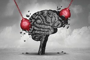
Concussions are among the most common neurological conditions, representing a substantial burden to adults and children.1 Persistent post-concussion symptoms (PPCS) are defined as symptoms that persist beyond the expected clinical recovery time frame: >10-14 days in adults and > 28 days in children.2 PPCS may include: nausea, dizziness, headaches, blurred vision, poor sleep, auditory disturbances, reduced executive function and emotional liability.1
Currently, research supports an active, symptom-based return to activity for individuals recovering from concussion. The Berlin consensus statement recommends 24-48 hours of rest before gradual return to activities.2 Active treatments may include subsymptom threshold aerobic exercise, cervical, vestibulo-ocular and cognitive therapies.3
Background
Two females ages 24 and 34, respectively, were referred to me from other health care providers for further assessment and management of their PPCS, which remained refractory to previous intervention. Previous management included: vestibulo-ocular rehab, aerobic exercise, psychotherapy, vision therapy, manual therapy for the cervical spinal, acupuncture, medication and suboccipital nerve blocks.
Case #1
A 24-year-old female enrolled in teachers college presented with PPCS of 14-months duration. She was injured after being elbowed in the head while falling out of a towable water tube during summer vacation.
At the time of presentation, previous treatment included vestibular rehabilitation, suboccipital nerve blocks, vision therapy, cervical manual therapy, and occupational therapy. Previous diagnostics included: MR imaging, CT scan, radiographs, and blood panel, all of which were interpreted as normal.
Her symptom burden at initial assessment included significant anxiety, particularly with visual motion and egocentric motion, dizziness, social irritability, poor short-term memory, left-sided tinnitus, neck pain, frontal and suboccipital headaches, motion sickness, and sensitivity to light and sound. She scored 54 on RPQ, 90 on PCSS and 16 on PHQ-9.
Physical examination was completed and revealed a pleasantly interactive woman with good attention and focus. Pulse 97, right-sided BP 133/83, left-sided BP 135/84; oxygen saturation 99%.
Gait was grossly normal. Upper and lower extremity light and sharp touch, as well as joint position sense, were normal. Pathological reflexes were absent. Reflexes were 2+ bilaterally at all levels. No evidence of pyramidal paresis, atrophy, flaccidity, spasticity or motor spontaneity noted.
Infrared video goggle (VOG) assessment revealed downbeat nystagmus with vision occluded. This was present in all seated head positions (head right, left, up, down, and tilted). Downbeat nystagmus was also produced in all directions of gaze eccentricity (right, left, up, down).
Bedside assessment of smooth pursuits revealed saccadic intrusions in left and down directions. Optokinetic gain was reduced in up and left directions. Choice reaction time was slow. Left head impulse testing produced saccadic refixations. Antisaccade testing showed 20 percent error rate and produced significant anxiety; testing also produced an increase in headache.
mCTSIB balance testing produced significant sway, which required assistance to prevent falls in head neutral, right, left and up conditions. Cervical spine joint position error was significant in all directions.
Other physical examination procedures including cranial nerve assessment, muscle tone, tandem stance, tandem gait, dual task gait, cervical orthopedic testing were unremarkable.
Case #2
A 34-year-old family physician presented with PPCS of 22 months duration. She was injured when a contractor's ladder fell on her head as she was leaving her home. The ladder knocked her to the ground and she lost consciousness for approximately three minutes.
At the time of presentation, previous treatment included vision therapy, cognitive behavioural therapy, psychotherapy, cervical manual therapy, suboccipital nerve blocks, neurofeedback, aerobic training, and clonazepam. Previous diagnostics included MR imaging, CT scan, radiographs, ECG, and blood panel, all of which were unremarkable.
Her symptom burden on initial assessment included significant brain fog and irritability, dizziness, neck pain, headaches, emotional liability, poor concentration, lightheadedness with orthostatic change and depersonalization. She scored 58 on RPQ, 100 on PCSS and 16 on PHQ-9.
Physical examination was completed and revealed a pleasantly interactive woman with good attention. Pulse 88, seated right-sided BP 118/71, left-sided BP 116/68, oxygen saturation 98%.
Gait was grossly normal. Upper and lower extremity light and sharp touch, as well as joint position sense, were normal. Pathological reflexes were absent. Reflexes were 2+ bilaterally at all levels. No evidence of pyramidal paresis, atrophy, flaccidity, spasticity or motor spontaneity noted.
Orthostatic vital sign assessment was completed; findings were as follows: five minutes supine HR and BP: 62 bpm and 118/70 mmHg; three minutes standing HR and BP: 79 bpm and 124/72 mmHg; 7 minutes standing HR and BP: 107 bpm and 121/74 mmHg.
During the seven minutes standing, the patient expressed an increase in headache and excessive emotionality. These values met the clinical criteria for the diagnosis of postural orthostatic tachycardia syndrome (POTS). Infrared VOG assessment revealed horizontal saccadic intrusions and convergence spasms with vision occluded. Bedside near-point convergence revealed divergence spasm at 8 and 11 cm. Antisaccade error rate was 30 percent with long latencies.
Optokinetic gain was reduced in all directions; following the stimulus, the patient felt significant dizziness. Luria's three-step test was abnormal on left. Rapid alternating movement testing revealed apraxia on the left. Vertical gaze holding produced eye pain and patient was unable to maintain fixation.
mCTSIB balance testing revealed significant sway in the medial-lateral direction, which was exacerbated in head up and left conditions. Other physical examination procedures including cranial nerve assessment, muscle tone, head impulse testing, vestibulo-ocular reflex exam, tandem stance, tandem gait, dual task gait, cervical orthopedic testing were unremarkable.
Multisystem Management Approach
In case #1, I made a clinical diagnosis of persistent post-concussion symptoms with vestibulo-oculomotor dysfunction, motor coordination impairment and cervical spine musculoskeletal impairments. Case #2 was diagnosed with persistent post-concussion symptoms and a clinical diagnosis of POTS.
Each patient was educated about PPCS and case #2 was educated about the clinical POTS diagnosis and that this should ideally be made through passive tilt table testing and medical evaluation. Management strategies were offered and both patients decided to participate in an individualized, targeted, multisystem neurorehab program.
Both patients received targeted treatment that matched their clinical dysfunctions. These strategies included: gaze stabilization training, ocular-movement exercises (smooth pursuits, saccades, OKN, convergence), spinal and extremity manipulation, motor coordination interventions, repetitive peripheral nerve stimulation, somatic sensorimotor complex movements, isometric contractions, and diaphragmatic breathing.
Each case had a unique element of treatment: Case #1 performed whole body on axis rotations with simultaneous VOR cancellation. Case #2 began treatment supine and was gradually elevated until her HR increased 10 bpm, at which point she was lowered 10 degrees and the treatment previously mentioned was performed.
Outcomes and Clinical Pearls
Both patients demonstrated significant improvements in their PPCS symptoms as measured by the PCSS, RPQ and PHQ-9. Following 15 consultations, case #1 reported a score of 2 on PCSS, 2 on RPQ and 0 on PHQ-9. She also reported complete resolution of all subjective complaints.
Following 12 consultations, case #2 reported a score of 6 on PCSS, 4 on RPQ and 2 on PHQ-9. Upon standing during orthostatic vital sign assessment, her HR increased 10 bpm initially and returned to supine resting levels within two minutes. She also reported significant improvements in her brain fog, irritability, emotional liability, lightheadedness and depersonalization.
This short case series demonstrates subjective and objective improvements in the symptoms and function of two females with PPCS following a multisystem, individualized and targeted rehab program to address their specific dysfunctions.
References
Polinder S, Cnossen MC, Real RG, et al. A multidimensional approach to post-concussion symptoms in mild traumatic brain injury. Front Neurol, 2018;9:1113.
McCrory P, Meeuwisse W, Dvorak J, et al. Consensus statement on concussion in sport—the 5th international conference on concussion in sport held in Berlin, October 2016. Brit J Sports Med, 2017;51(11):838-847.
Schneider KJ, Iverson GI, Emery CA, et al. The effects of rest and treatment following sport-related concussion: a systematic review of the literature. Brit J Sports Med, 2013;47(5), 304-307.
Building on a historic March 2026 meeting between Make America Healthy Again and chiropractic leadership, MAHA has announced the launch of the MAHA Chiropractic Hub, “a coordinated national partnership uniting MAHA Center, MAHA Action, and the chiropractic profession, including national associations, state organizations, practitioners, educators, researchers, and patient advocates. The Chiropractic Hub will advance federal policy, expand patient access, and build broad public support for chiropractic care across America.”
The chiropractic profession is confronting one of the most significant federal regulatory threats it has faced in decades. A new U.S. Department of Education (ED) accountability framework – commonly referred to as the “Do No Harm” (DNH) regulation – could place many chiropractic programs at risk of losing access to federal financial aid (student loans), potentially reshaping the future of chiropractic education and workforce development across the United States.
Pain has become the dominant language of musculoskeletal healthcare. Numeric pain-rating scales and symptom reports are routinely used as primary indicators of clinical success. But while pain reduction is meaningful, it is an incomplete and often misleading representation of recovery. This has real consequences for patient adherence, long-term outcomes, and how conservative care is perceived within the broader healthcare system.