It’s a new year and many chiropractors are evaluating what will enhance their respective practices, particularly as it relates to their bottom line. One of the most common questions I get is: “Do I need to be credentialed to bill insurance, and what are the best plans to join?” It’s a loaded question – but one every DC ponders. Whether you're already in-network or pondering whether to join, here's what you need to know.
Thoracic Pain: Tough Cases (Pt. 1)
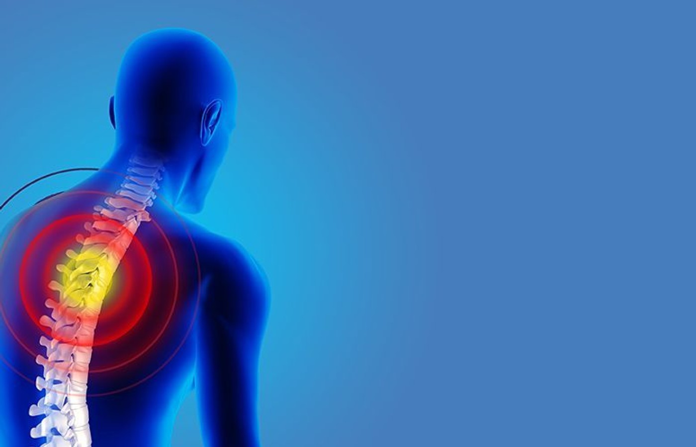
I have had a series of severe thoracic pain cases recently. These are not your typical "threw my rib out" patients. These folks have severe, chronic thoracic pain at the center of their lives, ongoing for months to years. Unlike the cervical or lumbar spine, there are rarely clinically significant anatomic pathologies to be identified.
My experience with chronic musculoskeletal pain tells me there is rarely a single contributor to the pain. If you are adjusting the same place, over and over, you are missing something. What are the factors that can contribute to this pattern? Let's focus on soft tissue, nerves and structure. Yes, there are biopsychosocial components that can contribute to any ongoing experience of pain. Once the patient is in the realm of chronic pain, we are dealing with some degree of central sensitization. For the purpose of this discussion, I'll assume you've already considered the obvious: spinal joints / thoracic segments.
1. The Ribs – From Back to Front
The anterior rib cage and the intercostals are so often ignored, and yet so important. Consider the posterior rib cage, the lateral rib cage and the anterior rib cage. In the posterior rib cage, you have the joints where the ribs meet the spine, as well as the region where the scapula glides over the posterior rib cage. The lateral rib cage technically has no joints. You are addressing the intercostal muscles, fascia and the intercostal nerves. Anteriorly, you have the sternochondral joints, as well as the anterior cartilage of the ribs, the anterior intercostals, and the muscles and fascia that overlie the chest.
2. Nerves That May Be Sensitized

Relevant nerves include the dorsal rami coming laterally from the thoracic spine, the dorsal scapular nerve coming through the scalenes from the lower cervical spine, and the intercostal nerves. You may treat these directly through gentle neuromobilization.1 Releasing the fascia and joints surrounding the nerves obviously impacts nerves. Don't assume your fascial release or adjustment has automatically downregulated the nerves. In chronic pain, the nerves themselves seem to become chronically inflamed pain generators.2
3. Additional Spinal Joints in the Lower Cervical Spine
Lower cervical dysfunction or lower cervical nerve irritation commonly refers pain (probably through the dorsal scapular nerve) into the upper thoracics. Palpate the segmental motion in multiple directions. Assess for a lower cervical segment resisting flexion and lateral bending; what could be called an "anterior cervical." The anterior lateral cervical fascia often feels tight and "ropy" over the scalene region.
4. Joints in the Shoulder
I tend to find frequent restriction of the sternoclavicular. It's worth checking the AC joint and the glenohumeral joint as well. The most common dysfunctional "shoulder joint" is likely to be the relation of the scapula to the rib cage.
5. Breathing and the Diaphragm
Does the patient predominately breath into their chest and/or neck? Even in those who move the respiratory diaphragm properly, they may not expand the rib cage in a lateral direction. Compare the side of pain to the other.
Below the anterior costal margin of the diaphragm, you'll find significant soft tissues. The inferior surface of the diaphragm is accessible to us via palpation. I frequently find tight fascial knots or bands in the diaphragm. Explore this entire area, lateral to medial.
Editor's Note: Part 2 of this article appears in the May issue and discusses other considerations when dealing with difficult cases of thoracic pain, including posture, scapular motion, muscular / fascial knots and viscerosomatic referral; as well as other considerations and how to approach working on sensitive areas such as the chest.
References
- "Yank Away Pain" – an approach focusing on irritated peripheral nerves.
- Boehnke H. "Dorsal Scapular Nerve Syndrome." Applied kinesiology approach.


