It’s a new year and many chiropractors are evaluating what will enhance their respective practices, particularly as it relates to their bottom line. One of the most common questions I get is: “Do I need to be credentialed to bill insurance, and what are the best plans to join?” It’s a loaded question – but one every DC ponders. Whether you're already in-network or pondering whether to join, here's what you need to know.
Shin Splints in Snowboarders
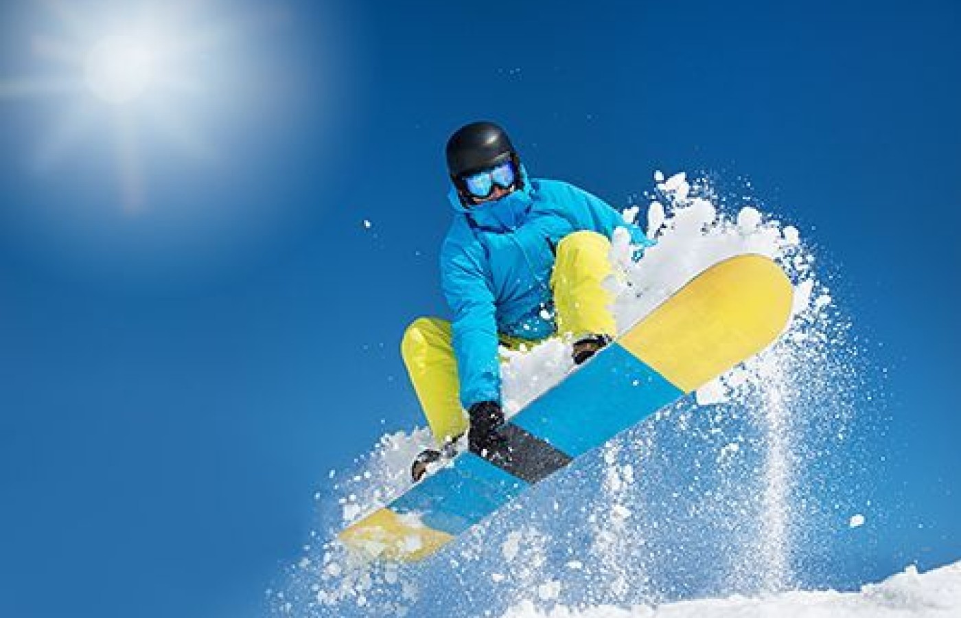
This time of year, snowboarders can be seen surfing powder, getting "big air" from jumps, and sliding on and off thin, metal rails at snow / ski resorts nationwide. Many people are captivated by the thrills they see on the slopes and on television, and decide to learn to snowboard themselves.
The first lesson involves learning to slide and stop on the heel-side edge of the board for steering and control. If the beginner lets their toe-side edge get too low, it catches in the snow and they are quickly slammed face first into the snow.
After several such incidents, riders learn to use their anterior tibialis muscles to keep the toe-side edge from catching. Most beginners are not accustomed to using these muscles, which can be strained within hours of snowboard riding.
Snowboarders aren't the only ones who can experience this problem. Runners also commonly strain the anterior tibialis when running downhill. Shin splints is the term used for a complaint of pain in the front of the calf. The eccentric stress of running downhill or snowboarding can create tissue damage at the tendinous insertions, fascia and muscle belly fibers.

The anterior tibialis muscle originates from the anterior surface of the lateral superior tibia and inserts into the medial cuneiform and first metatarsal bones of the foot. It is responsible for dorsiflexing and inverting the foot.
Diagnosis
Physical exam findings include tenderness and weakness of the anterior tibialis muscle. The pain may be exacerbated with stop-and-go running drills and/or walking downhill. When the anterior tibialis muscle is strained or irritated, it can cause pain, weakness and nerve compression, leading to tingling and numbness in the top of the foot and ankle.
Look for a loss of range of motion and/or loss of coordination with foot dorsiflexion. A dysfunctional anterior tibialis muscle can restrict ankle flexion, preventing a deep squat from being fully performed; and may also result in a foot drop with a "slapping" gait.
Muscle test bilaterally and note the deficiency of the involved muscle. Eccentric-break manual muscle testing involves having the patient lie supine with the foot fully dorsiflexed. Stabilize the lower leg with one hand and pull the patient's dorsiflexed foot downward.
Treatment
Effective correction of anterior tibialis dysfunction involves reducing the hypertonic state of the muscle. Thrusting and/or pressing into the tender insertion points will affect the nerve receptors located there. Using 2-3 gentle thrusts with your hands or an adjusting instrument should create enough stimulation to cause a significant improvement in muscle power output, pain reduction and increased mobility.
Adhesions in the muscle can be addressed by pressing into the tender muscle belly fibers and holding for 3-5 seconds. In my experience, this typically unlocks the fibers and restores greater mobility and function of the muscle. Post-treatment evaluation should be performed to determine treatment success and reveal whether further correction is needed.
Rehabilitation
The anterior tibialis can be stretched by opening the ankle joint. Instruct the patient to gently bend the toes and top surface of the foot against the ground into plantar flexion and then slowly roll the heel from side to side for five repetitions.
- Patient position: supine with foot dorsiflexed
- Doctor stance: foot of table
- Contact hand: top of forefoot
- Stabilization hand: lower leg
- Line of drive: pull foot inferiorly
Strengthening the anterior tibialis can be achieved by elevating the distal foot while standing on the edge of a step. I prefer that my patients work the good side first and then train the involved side. This can be done using 10-second isometric holds, multiple sets and reps, without resistance. Exercise tubing can be added to increase resistance.


