Evaluating and Correcting Sacral Misalignments: A Motion Palpation Approach
Russ Kalen, DC, CST
Editor's Note: Dr. Kalen addressed simplified adjusting of the pelvis in the Oct. 15 issue.
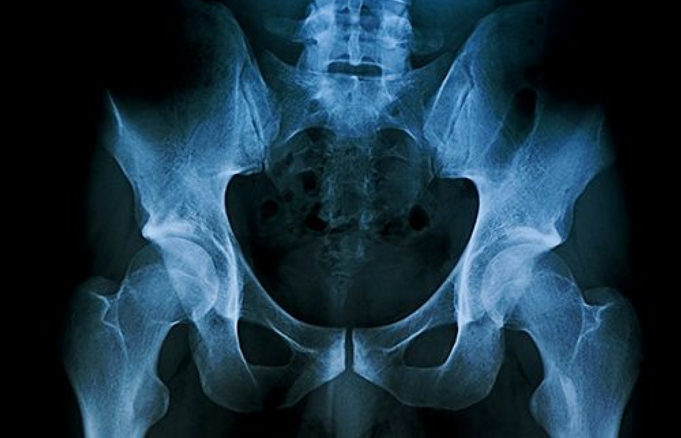
The movements of the sacrum within the pelvis are complex and often overlooked in chiropractic care. They are difficult to differentiate from other sacroiliac joint problems; however, once the pelvis is aligned, the remaining restrictions of the sacrum and coccyx are more straightforward to assess. In my previous article, I described my method of pelvic alignment. It starts with aligning the pubic symphysis and then each sacroiliac joint until the pelvis appears symmetrical in both alignment and freedom of movement. Now let's discuss how to evaluate and correct sacral misalignments.
Evaluation Techniques
The sacrum can be readily evaluated using the craniosacral rhythm (CSR). This method of palpation uses the inherent motion of the sacrum without disturbing the surrounding structures, so you can discern the direction and ease of motion of the sacrum independent of the pelvis. For those of you who are unfamiliar with craniosacral therapy, the CSR is a rhythm created by the pressure of the cerebrospinal fluid as it circulates through the brain and spinal cord.1 For our purposes here, understand that this rhythm creates a regular flexion and extension of the sacrum of about 6 mm to 12 mm of ROM over a 10-second period.
(If you look to the literature, you will find all manner of descriptions and rates for the CSR, many of them more poetic than scientific.2 In my frustration with this state of affairs, I have been measuring and documenting the CSR in all my patients since 1990. In my experience, healthy patients over the age of 8 all had a CSR that measured 10 seconds a cycle +/- 0.5 seconds.)
To palpate the motion of the sacrum, sit beside the patient facing their feet. Place your hand on the sacrum with the fingertips at the coccyx. This position is chosen so your hand is free to side-bend and rotate easily along with the sacrum.
Relax and follow the sacrum through its flexion and extension, trying not to induce motions. As the sacrum moves, it is possible to evaluate for loss of motion or asymmetrical motion, such as side-bending on flexion or rotation about the long axis. It also should be noted that the method of craniosacral “listening” is different than many other methods of palpation, in that you are not inducing a motion, but merely palpating an existing motion. Because of this, it is important for you to remain relaxed; otherwise, you will only be aware of your own muscle tension. The motion is quite visible when your hand is following the sacrum.
Findings and Corrections
Reduced sacral gliding: The most common finding is that the sacrum will not glide smoothly on one side. This creates a rubbing or vibration that can be felt to one side of your hand and is likely due to residual tension in the sacroiliac ligaments within the joint. You need only to determine whether it is the flexion or extension resisting the movement and create an adjustment to the sacrum that assists the direction of reduced movement.
L5 anterior to S1: Another common finding is when the sacrum runs into a hard endpoint after a short excursion in the extension direction. This usually indicates L5 is anterior to S1. My preferred adjustment for this is an anterior-to-posterior drop-table adjustment in which the patient is positioned supine with the pelvis on the drop mechanism and L5 just above it.
Place a support under the sacrum (a folded hand towel works well) to isolate the sacrum from the pelvis so the sacroiliac joints are not affected by the drop table. Then place a palm over the lower abdomen to help focus the drop to the L5 – S1 junction and bring the patient's knees to the chest. The flexion uses the tension of the lumbosacral ligaments to help draw L5 posterior. The drop is initiated by your pressure on the legs only. Of course, if there is a bilateral pars defect of L5, the flexion is less important.
Lumbar / sacral nerve involvement: The nerves of the lumbar and sacral plexus normally are able to slide in and out of the IVF and sacral foramina with the movements of the lower extremity.3 When they don't, the sacrum and coccyx will have a restricted or lateral movement on flexion, or the coccyx will move in extension when the sacrum moves into flexion. This suggests the filum terminale is under tension from either the lumbar or sacral nerves on the same side.
To find the fixation, induce a slight flexion of the coccyx and then listen with the other hand over the lower spine to feel where the motion is blocked. Adjusting the appropriate lumbar segments often will relieve the problem and can be evaluated immediately by again palpating the sacral coccygeal motion. Rarely, the nerve plexus may be restricted by loss of nerve glide in the fascia of the ipsilateral leg.
When the tension is in the sacral plexus, it will feel as though pressure increases under your hand during flexion. In this case, mobilization of the fascia through the involved sacral foramina often will free the nerve glide and restore the sacral flexion. For this treatment, the patient remains prone on the adjusting bench while you place your hand palm up under their lower abdomen (above the pubis) and press toward the anterior sacrum. The other hand contacts the fascia over the sacral foramina and the pressure is downward into the foramina. The alternate hand pressures to mobilize the fascia back and forth through the foramina several times. This attempts to restore normal nerve mobility through the foramen.3 You can recheck the sacral mobility in flexion to see if the correction is successful.
Other Considerations
These treatments are powerful and may induce a temporary increase in inflammation, so the patient should be advised to manage this reaction over the following two days with appropriate anti-inflammatories as necessary. My experience is the patient usually has more ease of movement upon leaving the office and then a few hours later, may feel sore in a new way. This reaction usually lasts for 24 hours, after which time the patient is markedly better.
As I noted in my previous article, patients with hemivertebrae, spinal fusion, laminectomies and other such structural anomalies should be treated cautiously, as the corrections achieved may be greater than what the structures can sustain. Many of these treatments directly remove pressure from nerves in the lumbar or sacral plexus, thus treating various types of sciatica, heel pain, numbness and other conditions that involve symptoms in the lower extremities. In my experience, freeing the lower spine in this way also may affect conditions such as migraine and TMJ dysfunction. This is the magic of chiropractic.
References
Upledger J, Vredevoogd J. Craniosacral Therapy. Seattle, WA: Eastland Press, 1983, pp. 6-13.
Sills F. Craniosacral Biodynamics. Berkeley, CA: North Atlantic Books, 2001, pp. 38-394.
Haldeman S. Modern Developments in the Principles and Practice of Chiropractic. New York, NY: Appleton-Century-Crofts, 1980, p. 49.
Building on a historic March 2026 meeting between Make America Healthy Again and chiropractic leadership, MAHA has announced the launch of the MAHA Chiropractic Hub, “a coordinated national partnership uniting MAHA Center, MAHA Action, and the chiropractic profession, including national associations, state organizations, practitioners, educators, researchers, and patient advocates. The Chiropractic Hub will advance federal policy, expand patient access, and build broad public support for chiropractic care across America.”
The chiropractic profession is confronting one of the most significant federal regulatory threats it has faced in decades. A new U.S. Department of Education (ED) accountability framework – commonly referred to as the “Do No Harm” (DNH) regulation – could place many chiropractic programs at risk of losing access to federal financial aid (student loans), potentially reshaping the future of chiropractic education and workforce development across the United States.
Pain has become the dominant language of musculoskeletal healthcare. Numeric pain-rating scales and symptom reports are routinely used as primary indicators of clinical success. But while pain reduction is meaningful, it is an incomplete and often misleading representation of recovery. This has real consequences for patient adherence, long-term outcomes, and how conservative care is perceived within the broader healthcare system.