New York's highest court of appeals has held that no-fault insurers cannot deny no-fault benefits where they unilaterally determine that a provider has committed misconduct based upon alleged fraudulent conduct. The Court held that this authority belongs solely to state regulators, specifically New York's Board of Regents, which oversees professional licensing and discipline. This follows a similar recent ruling in Florida reported in this publication.
The Struggle to Heal the Cervical Syndrome: Exploring Challenges and Solutions
Editor's note: This is the first article in a series on chiropractic techniques to treat challenging patient presentations.
Chiropractors of earlier times were reliant on manipulation only, and some conditions we treat today must be primarily manipulation-based. Of course, ancillary procedures will only aid in recovery, but in the struggle to heal the cervical syndrome, only manipulation of the right kind and in the right amount will result in a healthy, happy patient.
Symptom Portrait
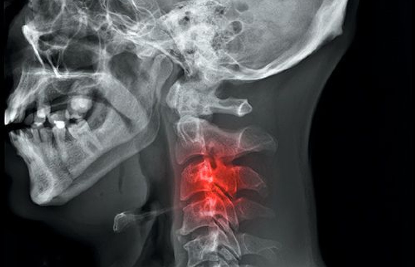
Based on our Google and Amazon searches, Ruth Jackson, MD, published The Cervical Syndrome in 1958, although we did find a review of a 1956 publication. The cause of the symptoms was a bit nebulous, but was based on postural and osteologic changes noted on X-ray. However, clinically, we can see the same symptoms in most age groups before any bone changes are noted on X-ray.
This is a quote from Mobilisation of the Nervous System (pp. 44-45), by David Butler, 1991: "Clinically, apparent mechanical impairment of the sympathetic chain is often evident. Perhaps this provides an explanation for symptoms such as nausea, vague thoracic pains and headaches evoked by a SLR. Slump tests occasionally reproduce odd symptoms such as deep abdominal pains, flushes and sweating. Upper Limb Tension Tests can cause a ‘pumping feeling' in the arm and symptoms of increased sweating and color changes in the extremity are often related to limbs with positive tension tests." He also describes "adverse mechanical tension on the sympathetic nervous system as the cause of such impairments."

A common misconception is that nerve-root irritation from disc protrusion is due to compression. However, neurologically compression causes paresthesia or anesthesia. The pain of disc protrusion is due to the stretch of the nerve and inflammatory components. Similarly, increased mechanical tension of the sympathetic nervous system is what causes increased firing of those nerves. The sympathetic nervous system is also the most exposed and least protected of any of the separate parts of the entire nervous system.
The simplest explanation of the cervical syndrome is that it is an up-regulation of the sympathetic nervous system. The sympathetic nervous system should be on standby only for "fight or flight" situations. The parasympathetic nervous system should be predominant in "rest and digest" mode. This is the healing environment the human body needs.
The symptoms of the cervical syndrome are easy to predict and include any symptoms that are the opposite of "rest and digest." The following list is from Len Faye, DC, which he provides to his patients. Symptoms include, but are not limited to:
- Headache that usually starts in the back of the neck or base of the skull and radiates to the head or around the ears
- Slight blurring of vision with slight pupil dilation
- Dizziness or easy loss of balance
- Nausea or poor digestion
- Pressure behind the eyes
- Unexplained fainting attacks
- Ringing in the ears
- Numbness in side of the face, neck, head or tongue
- Tight neck muscles
- Difficulty swallowing
- Pain behind shoulder blade
- Shoulder pain, arm pain, tennis elbow or carpal tunnel syndrome
- Weakness, pain, or tingling in arm, forearm and/or hand; weak grip
- Increased sweating
- Shortness of breath
- Heart palpitations, slightly elevated blood pressure or fast heart rate
- Frontal chest pain
- Numbness and tingling of the fingers when awakening
- Anxiety, panic attacks or insomnia
Most of these symptoms are neurological in nature, but a few are purely postural, such as difficulty swallowing. Frontal chest pain or pseudo-angina can be attributed to scalene trigger points, as noted in Travell and Simons' Myofascial Pain and Dysfunction: The Trigger Point Manual.
We should probably include the common postural changes that cause up-regulation of the sympathetic nervous system. The first and most obvious is the protracted shoulder / forward-head posture. This causes an upper thoracic flexion posture with the inability to extend the upper thoracic spine. This restriction is the most difficult and also most incorrectly treated area in the entire spine.
The second part of the postural dysfunction is the lower cervical spine. This will probably generate some debate, but it has to be said. Cervical hypolordosis as we see on so many X-ray reports is probably a myth, about 5 percent of the population being the exception. Most lower cervical spine postures are actually hyperlordotic. In Atlas of Normal Radiographic Anatomy and Variants That May Stimulate Disease, by Keats and Anderson, it is noted that most cervical hypolordosis on film is based on patient positioning.
We challenge practitioners to look at the patient and not the film. If the cervical spine were hyperlordotic, it would appear as a skin crease just like it appears in spondylolisthesis of the lumbar spine. The debate is based on where the adaptation occurs due to the increased upper thoracic flexion. It can occur anywhere in the cervical spine. We believe it occurs at the lower cervical region.
Chiropractic Treatment Options
Treatment of the cervical syndrome is moderately simple. Let's consider a particular type of patient. Today's patient is a hyperflexible patient – the most difficult. If you are not analyzing your patients' flexibility, you are increasing your chances for failure. Two methods will help the doctor understand which patient type has entered the office: Harrington's and Beighton's criteria. A quick Google search will have you competent in these in five minutes or less.
Each adjustment should always be based on movement loss, patient's age, gender, flexibility and chronicity. Often, doctors use only their favorite technique system, which is great for some patients, but horrible for others. As an example, I love seated cervicals, but they are a horrible choice for the hyperflexible. They will still work, but the force required to achieve cavitation is too aggressive when another method will suffice with less force.
Although every case is different and should be based on your palpation, the common elements of this condition are a lack of upper thoracic extension, a lack of lower cervical flexion and anterior-to-posterior rotation, and sternoclavicular depression and retraction. Here is a basic outline of a few of the possible palpation / adjustment protocols.
When you review the demonstration videos for these protocols [accessible by viewing the app version of this article], note that the upper thoracic spine adjustments are performed supine. The reason is that typically, prone upper thoracic manipulation creates upper thoracic flexion, which is not easily forced into extension without massive doses of force.
Cervicothoracic Palpation and Decision-Making:
- Observe posture: hyperkyphotic = hyperflexed = inability to extend; hypokyphotic = hyperextended = inability to flex.
- Use protraction-retraction to determine the need for a flexion or extension adjustment.
- Have the seated patient roll the chin forward (protraction) while your thumb is between the spinous processes – a gapping will occur unless they lack flexion.
- Have the patient return to neutral and then a retract a little – the spinous processes should push your thumbs out of the gap unless the patient lacks extension.
- Use the coupled rotation palpation to determine laterality (side) of fixation – although most actually are bilateral.
- Stabilize the head with one hand on top of the head; use the other hand to palpate with the thumb on the lamina of C7 though T4.
- Slightly flex the cervical spine and rotate and laterally flex – also slight – and challenge the joint from posterior to anterior.
- With the adjustment, first satisfy the sagittal axis component and then as it improves, begin to add coupled rotation and lateral flexion.
Upper Thoracic Extension Manipulation:
- Patient supine with arms crossed and hands under the axilla; from the right side of the table, the left arm would be on top.
- Roll the patient toward you and make contact with the right hand.
- The contact hand is a raised thenar contact; the tip of your thumb and the base of the base of the third and fourth metacarpalphalangeal joints.
- The contact point on the patient is the left lamina with a tissue pull caudally.
- Contact the patient's right arm at the elbow and begin to roll them onto the contact, while contacting their right arm with the side of your chest.
- Roll the patient approximately 30 degrees onto the contact and perform a slight drop (don't overdue the force; it will move easily).
Index @ 90 Degrees for Lower Cervical AP Rotation Restrictions:
- Patient is supine; doctor is perpendicular to the table and well over the patient.
- Indifferent hand – contact the zygomatic process with thenar pad and the mastoid with the middle digit.
- Rotate the patient's head a full 90 degrees and let your indifferent hand sink into the headpiece of the table (this manipulation cannot be used on patients who cannot rotate fully).
- Use a very light index contact at the metacarpalphalangeal joint on the transverse process of the restriction (usually C5 or C6).
- Apply very light pressure on the contact while very slightly lifting the head.
- Add a very quick, short impulse thrust into the table.
- This procedure will take time to master – it has a difficulty scale of 10 out of 10.
Upper Thoracic Mobilization
- Patient is placed supine over a half foam roll and brought down the table until their head if off the table significantly. The easiest way to do this is bring them off the table until their arms clear the table – the axillas will be at the edge of the table.
- The arms hang free, opening up the pectoralis muscles and the sternoclavicular joint.
- The patient's palms should be facing the ceiling.
- The patient's head is supported with one hand under the occiput and the other on the chin, with the tip of the chin splitting the third and fourth digits.
- At this point, have the patient squeeze the table by straddling the table with their legs and feet.
- A strong traction is applied – their whole body will move; then retract the cervical spine with the third and fourth digit contact on the chin. The patient's cervical spine will be fully extended to their limit.
- Repeat process 10-15 times. During the process, talk to the patient and ask if they feel tension elsewhere in their spine. If they do, stop the process, palpate and manipulate that area, and begin again.
- Contraindicated if patient is dizzy.
- The procedure is usually well-tolerated, but has little effect on extremely flexible people.
- Clinical tip – very effective for adhesive capsulitis, as we are witness to personally!
The last piece of advice on this condition is to allow time for change to occur. The principle of SAID (Specific Adaptation to Imposed Demands) has to apply. Again, this principle is from Len Faye, DC. We don't seem to have a gray area in terms of treatment schedules; we either have too many or too few. The insurance companies cannot dictate treatment schedules; the patient's biomechanical needs must be foremost, particularly when treating the cervical syndrome.


