It’s a new year and many chiropractors are evaluating what will enhance their respective practices, particularly as it relates to their bottom line. One of the most common questions I get is: “Do I need to be credentialed to bill insurance, and what are the best plans to join?” It’s a loaded question – but one every DC ponders. Whether you're already in-network or pondering whether to join, here's what you need to know.
Achilles Injuries, Part 1: Insertional Tendinitis
Despite its broad width and significant strength, the Achilles tendon is injured with surprising regularity. In a study of 69 military cadets participating in a six-week basic-training program (which included distance running), 10 of the 69 trainees suffered an Achilles tendon overuse injury.1 The prevalence of this injury is easy to understand when you consider the tremendous strain runners place on this tendon. For example, during the push-off phase of the running cycle, the Achilles tendon is exposed to a force seven times average body weight. This is close to the maximum strain the tendon can tolerate without rupturing.2 And when you couple the high strain forces with the fact that the Achilles tendon significantly weakens as we get older, it is easy to see why this tendon is injured so frequently.
Anatomically, the Achilles tendon represents the conjoined tendons of the gastrocnemius and soleus muscles. Although the plantaris muscle runs between the gastrocnemius and soleus, it is technically not considered part of the Achilles tendon. Approximately 5 inches above the Achilles attachment to the back of the heel, the tendons from gastroc and soleus unite to form a single thick Achilles tendon. These conjoined tendons are wrapped by a single layer of cells called the paratenon. This sheath-like envelope is rich in blood vessels necessary to nourish the tendon.

The tendon itself is made primarily from type-1 and type-3 collagen. In a healthy Achilles tendon, 95 percent of the collagen (which accounts for 70 percent of the dry weight of the tendon) is made from type-1 collagen, which is stronger and more flexible than type 3. It is the strong crosslinks and parallel arrangement of the type-1 collagen fibers that give the Achilles tendon its strength.
The Achilles tendon is unusual in that, at about the point where the gastroc and soleus muscles unite, the tendon suddenly begins to twist, rotating a full 90 degrees before it attaches to the back of the heel. (Figure 1) This extreme twisting significantly improves efficiency in running because it allows the tendon to function like a rubber band, absorbing the gravitational energy present during the early phases of the gait cycle and returning the energy in the form of elastic recoil during the propulsive period.
In fact, recent research confirms that the gastroc and soleus muscles perform relatively little work during the propulsive period. Rather than concentrically shortening to propel the body forward by forcefully plantarflexing the ankle with a strong concentric contraction, the gastrocsoleus muscles fire almost isometrically during late midstance, forcing the Achilles tendon itself to stretch and return energy.
The belief that muscles act merely to adjust tension on spring-like tendons was first demonstrated in a study by Roberts, et al.3 These researchers surgically implanted special crystals and strain gauges into the gastroc muscles and Achilles tendons of turkeys that were forced to run on treadmills. When the turkeys ran at faster speeds, the authors noted that the gastroc and soleus muscles maintained an almost fixed length, firing with a nearly isometric contraction during early propulsion. The "isometric impulse" of the gastrocsoleus complex allows the rotated Achilles tendon to unwind and immediately snap back, storing and returning energy at just the right moment.
By calculating muscle activity and force production, Roberts, et al., confirmed that the storage and recovery of energy in the stretched tendon was responsible for more than 60 percent of the work performed by the muscle-tendon complex. The results of this study make it clear that the spring-like action of the Achilles tendon acts to significantly lessen the metabolic expense of running by reducing the workload on the muscles (which consume calories, generate heat and require removal of waste products such as lactic acid).
By passively storing and returning energy, the Achilles tendon allows the leg to behave like a very efficient pogo stick. According to Anderson,4 optimizing the storage and return of energy requires considerable practice as it is a learned process, involving precise timing of numerous kinetic and kinematic variables.

Even though the storage and return of energy inside the Achilles tendon improve metabolic efficiency, the extreme forces stored in the tendon increase the likelihood of injury. Depending upon the location, Achilles injuries are divided into three categories: insertional tendinitis, paratenonitis and non-insertional tendinosis. Let's focus on the insertional injuries (paratenonitis and non-insertional tendinosis will be discussed in a subsequent article).
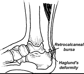
As the name implies, insertional tendinitis refers to inflammation at the attachment point of the Achilles on the heel. This type of Achilles injury is extremely difficult to treat and typically occurs in high-arched, inflexible individuals, particularly if they possess a Haglund's deformity. (Figure 2) Because a bursa is present near the Achilles attachment, it is common to have an insertional tendinitis with the retrocalcaneal bursitis.
Until recently, the perceived mechanism for the development of insertional tendinitis seemed straightforward: The individual possessing a high arch and tight gastrocnemius/soleus creates an increased tensile strain in the posterior aspect of the Achilles tendon when the ankle is maximally dorsiflexed during late midstance / early propulsion. Although logical, recent research has shown that just the opposite is true.5
By placing strain gauges inside different sections of six cadaveric Achilles tendons and loading the tendons with the ankle positioned in a variety of angles, researchers from the University of North Carolina discovered the posterior portion of the Achilles tendon is exposed to far greater amounts of strain (particularly as the ankle is dorsiflexed), while the anterior aspect of the tendon, which is the section most frequently damaged with insertional tendinitis, is exposed to very low loads.
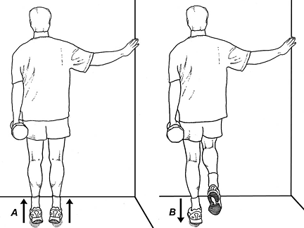
The researchers suggest that the lack of stress on the forward aspect of the Achilles tendon (which they referred to as a tension-shielding effect) may cause that section to weaken and eventually fail. As a result, the treatment of an Achilles insertional tendinitis should be to strengthen the anterior aspect of the tendon. This can be accomplished by performing the eccentric load exercise illustrated in Figure 3.
It is theorized that the slight vibration associated with the eccentric component of these exercises stimulates fibroblasts to repair the tendon.6 By raising up off your heels and slowly lowering yourself, you can feel the slight muscle vibration during the eccentric component of the exercise. It is particularly important to perform eccentric exercises starting with the ankle maximally plantarflexed, because this position places greater amounts of strain on the more frequently damaged anterior aspect of the tendon.
When lateral insertion injuries occur in individuals with cavovarus feet, an effective treatment protocol is to incorporate valgus wedges. These inexpensive wedges can be purchased from several distributors. Even though heel lifts are frequently prescribed to treat this injury, recent evidence suggests that when used on individuals with reduced ranges of ankle dorsiflexion, heel lifts actually increase EMG activity in the medial gastrocnemius and tibialis anterior muscles.7 Because this is consistent with prior research suggesting that heel lifts may increase strain on the Achilles tendon,8 the routine prescription of heel lifts for the treatment of Achilles tendon disorders needs to be reconsidered.
Rather than accommodating tightness in the gastrocsoleus complex with heel lifts, a better approach is to lengthen these muscles with gentle stretches. Because overly aggressive stretching can damage the insertion by pulling on it too vigorously, the stretches should be performed with mild tension and held for 35 seconds. If morning pain is present, a night brace should be considered.
To lengthen the gastrocsoleus muscles as quickly as possible, trigger points in the gastrocsoleus complex should be massaged vigorously before stretching. As demonstrated in several studies, stretching programs incorporating deep-tissue massage have been shown to increase range of motion more effectively.9-10 Remember that because the Achilles tendon rotates approximately 90 degrees, contracture in the lateral gastroc may produce a medial insertion injury and vice-versa.
In addition to incorporating massage to lengthen the muscles, deep-tissue massage should also be used on the tendon insertion. As verified by Loghmani and Warden,11 controlled microtrauma has been shown to stimulate repair by accelerating fibroblastic remodeling. This is in contrast to many common NSAIDs, such as indomethacin and celecoxib, which have been recently shown to significantly reduce tendon-to-bone healing in laboratory animals.12 Obviously, these drugs should be avoided in the management of insertional Achilles injuries.
References
- Mahieu NN, Witvruow E, Stevens V, et al. Intrinsic risk factors for the development of Achilles tendon overuse injuries. Am J Sp Med, 2006;34:226-35.
- Allenmark C. Partial Achilles tendon tears. Clin Sports Med, 1992;11:759-769.
- Roberts TJ, Marsh RL, Weyand PG, Taylor CR. Muscular force in running turkeys: the economy of minimizing work. Science, 1997;Feb(275):1113-15.
- Anderson T. Biomechanics and running economy. Sports Med, 1996;22:76-89.
- Lyman J, Weinhold P, Almekinders LC. Strain behavior of the distal Achilles tendon. Am J Sp Med,
- Rees J, Lichtwark G, Wolman R, et al. The mechanism for efficacy of eccentric loading an Achilles tendon injury: an in vivo study in humans. Rheumatology, 2008;47:1493-1497.
- Johanson M, Allen J, Matsumoto M, et al. Effects of heel lifts on plantarflexor and dorsiflexion activity during gait. Foot Ankle Int, 2010;31:1014-1020.
- Dixon S, Kerwin D. The influence of heel lift manipulation on Achilles tendon loading in running. J Appl Biomech, 1998 Nov;14(4).
- Hopper D, Deacon S, Das S, et al. Dynamic soft tissue mobilization increases hamstring flexibility in healthy male subjects. Br J Sports Med, 2005;39:594-598.
- Guler-Uysl F, Kozanoglu E. Comparison of the early response to two methods of rehabilitation for adhesive capsulitis. Swiss Med Wkly, 2004;134:363-368.
- Loghmani M, Warden S. Instrument-assisted cross-fiber massage accelerates knee ligament healing. J Orthop Sports Phys Ther, 2009;39:506-514.
- Cohen D, Kawamura S, Ehteshami J, Rodeo S. Indomethacin and celecoxib impair rotator cuff tendon-to-bone healing. Am J Sports Med, 2006;34:362-369.
Author's note: Part 2 of this article will discuss innovative treatments for the management of non-insertional Achilles injuries.


