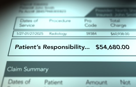
Recent laws in New Jersey and California represent a disturbing trend that will negatively impact a practice’s ability to collect monies from patients, as well as expose them to significant penalties if the practice does not follow the mandatory guidelines to a T. Please be aware that a similar law may be coming to your state. The time to act is before the law is passed.
The Straight Leg Raise Test
I recently had the chance to review a defense exam on an auto accident case. This was a classic rear-end whiplash type of injury, and the patient had pursued a normal course of chiropractic treatment. In reviewing the defense report, one of the interesting things I noted was the brief nature of the physical exam: some palpation, some observation of motion, and only one orthopedic test - the straight leg raise (SLR).
There is probably no more commonly used orthopedic test than the SLR. Almost every report I review indicates that the doctor performed the maneuver, even if there were no lower back complaint. The SLR is a great test, but it should not be used alone to arrive at a diagnosis.
Evans' text reminds us that the lumbar nerve roots have a narrow range of movement for stretching. The nerve roots are not brought to tension and stretched by the SLR until 35 to 70 degrees of angulation have been reached. If there is compromise of the normal space (i.e., disc bulge, inflammation) this space is used up and the pain will manifest more quickly - thus giving you a positive finding of pain. It is important to evaluate fully a patient with a positive SLR, as nerve root compression may mimic sacroiliac inflammation. When performing the SLR, remember that sciatic pain in the leg produced from 0 to 30 degrees indicates nerve root compression. Sciatica produced between 30 and 60 degrees indicates sacroiliac disease. Sciatic pain produced with leg motion beyond 60 degrees points to lumbosacral conditions.1,2
The classical SLR test is performed with the patient lying supine with the legs fully extended. (In the patient with lower back pain, the leg with pain is the one evaluated.) The examiner places one hand under the ankle of the affected leg and the other hand on the knee - and then lifts the ankle and flexes the thigh relative to the pelvis. The test is considered positive if pain is reproduced or increased in the lower back or leg. If the SLR is ever positive, further testing must be pursued to define the nature of the irritation. It has been my experience that most docs stop with just the SLR. However, there are a few more simple steps that can be added easily and will increase the diagnostic value of this maneuver:
Goldthwaite's Test: Slide your hand under the patient's lower back and feel the lumbosacral spinous processes. As you lift the leg to a point of pain, feel for motion between these segments. If pain is experienced before the spinous processes separate, this suggests the irritation is rooted in the sacroiliac joint. If the pain manifests with motion of the lumbar segment, the lesion is more likely in that area.
Lift the Head: Once the leg is raised to the point at which symptoms are reproduced, instruct the patient to lift his or her head, bringing the chin to the chest. If this movement is limited or increases the pain in the lower back or leg, it suggests inflammation of the nerve root.
Bragard's Sign: If the SLR is positive, lower the leg on the affected side to just below the point of pain and quickly dorsiflex the foot. If the pain is duplicated or increased, this suggests sciatic neuritis.
Sicard's Sign: If the SLR is positive, lower the leg to just below the point of pain and quickly dorsiflex the great toe. If the pain is duplicated or increased, this suggests sciatic radiculopathy.
Cox Sign: If the patient raises the affected hip off the table instead of flexing the hip, this indicates prolapse of the nucleus into the IVF.
Seated SLR (Lesegue Sitting Test): With the patient seated, the affected leg is raised to the point of pain. The test is considered positive if pain is reproduced or increased in the lower back or leg. To avoid pain in the leg, the patient may lean back; this also would be considered a positive finding. This test is also a good cross-screening for malingering, as pain with a SLR should be reproduced with the seated SLR.
Deyerle's Sign: With the patient seated, the affected leg is raised to the point of pain. The knee is then slightly flexed and pressure is applied into the popliteal fossa. If the radicular symptoms are increased, the test is positive for sciatic nerve irritation above the knee due to stretching of the nerve over an abnormal mechanical obstruction.
Well Leg Raise: The SLR is performed on the unaffected leg. If pain is referred back to the symptomatic side, this indicates nerve root compromise by an extruded disc.
Fajerstajn's (pronounced "fire-stines"): This test is the same as Bragard's, just performed on the unaffected side. Pain produced with this maneuver suggests IVD syndrome or dural adhesions.
Vleeming's Active SLR: This test was discussed in detail by Dr. Craig Liebenson in the Feb. 24, 2003 issue of DC. Basically, pain or poor motion control with active performance of the maneuver suggests SI joint dysfunction or compromised hip flexors.3
So, what have I learned? The SLR is a great test, but it should not stand alone. If your patient experiences pain with a SLR, you are obligated to test further to define the source of the patient's complaints. Remember that when performing the SLR, always note where the pain goes. Is it in the back, the buttock or the leg? Does it go down to the knee or foot? Does it cause tension or pulling up into the neck?4 Such notes are invaluable when documenting your patients' complaints and the extent of the irritation. These extra notes help document the severity of the patients' complaints and show the progressive response to care. This extra documentation can also help make the difference if you must justify your diagnosis to an insurer or third party. Take the extra few seconds to add these tests into your exam routine. They will serve you well.
References
- Evans, RC. Illustrated Essentials in Orthopedic Physical Assessment. St. Louis, Missouri: Mosby, 1994.
- Hoppenfeld, S. Physical Examination of the Spine and Extremities. San Mateo, CA: Appleton & Lange, 1976.
- Liebenson, C. Vleeming's active SLR test as a screen for lumbopelvic dysfunction. Dynamic Chiropractic, Feb. 24, 2003. www.chiroweb.com/archives/21/05/13.html.
- Hammer, W. Use of the straight leg test for upper extremity involvement. Dynamic Chiropractic, Nov. 17, 1997. www.chiroweb.com/archives/15/24/22.html.


