It’s a new year and many chiropractors are evaluating what will enhance their respective practices, particularly as it relates to their bottom line. One of the most common questions I get is: “Do I need to be credentialed to bill insurance, and what are the best plans to join?” It’s a loaded question – but one every DC ponders. Whether you're already in-network or pondering whether to join, here's what you need to know.
Identification and Treatment of Muscular Chains
The mechanism of injury of bones, ligaments, tendons and other soft tissues is tissue failure. Tension, shear, compression and torsion are the most likely types of stresses which produce tissue strain. Cumulative microstrain or traumatic macrostrain can both overload a tissue's capacity to resist buckling and failure. Whether or not injury occurs depends of course on external factors such as duration, intensity or frequency of exposure to biomechanical stress, but it also depends on the ability of the body to resist and control such forces so as to maintain stability. Stabilization is a result of the interaction of three subsystems: the CNS for control; the passive osteoligamentous structures for passive restraint and sensory feedback; and the active muscle system for production and control of movement.
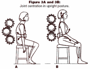
Most injuries occur as a result of repetitive end-range loading. Tissue protection occurs when agonist/antagonist muscles are coactivated to maintain control of tissues within the physiological range and avoid harmful end-range loading while maintaining the centrated position of joints. Joint centration is the point of equilibrium or maximum congruence for optimization of handling joint loading. The ability of muscles to achieve and maintain centration concerns both coordination and endurance functions.
To promote or restore joint stability, rehabilitation of the muscular system is necessary. This requires a holistic approach emphasizing the functional role of muscles in maintaining dynamic joint stability while producing the movements which are required by our activities of daily living. This functional role views muscles as working together in chains to perform functional activities, rather than as individual muscles having the classical roles expressed as their actions.
Types of Muscular Dysfunction
The muscular system can respond to trauma, repetitive overload, pathology or pain in a variety of ways. Muscles can become facilitated, overactive and even shortened in length, or they can become inhibited, weakened and atrophic. Individual muscles function in concert with entire groups of muscles throughout the body to maintain stability or produce movement. These individual muscles may be classified as agonists, synergists or antagonists. When any muscle's function is disturbed (i.e., facilitated or inhibited), movement and stability throughout the entire kinetic chain will be affected. Clinically, we see the three following situations. One: muscle imbalance where a movement agonist is inhibited while its antagonist is facilitated (+/-); two: agonist-antagonist facilitation (+/+); and three: agonist-antagonist inhibition (-/-).
Muscle imbalance is probably the most common clinical presentation. It occurs in a systematic fashion.1 Patients with muscle imbalance present with predictable shortening (decreased muscle length) in typical muscles such as the upper trapezius, suboccipitals, erector spinae, iliopsoas and hamstrings with concomitant lengthening or inhibition of the lower trapezius, deep neck flexors, deep abdominals and gluteals. Why is this so? Certain muscles which relate to the fetal position, static work postures or slumping have a tendency to become overactive or even shorten, while other muscles which relate to the neurodevelopment of upright posture or dynamic joint stability tend to become inhibited or even weak. Modern society's emphasis on constrained postures and sedentary lifestyles promotes an imbalance between overactive and inhibited muscles. The development of these predictable muscle imbalances is further spurred by the diminished afferent flow of sensory information from the periphery, in particular the sole of the foot, due to sedentarism and a lack of variety of movement. Naturally, movement patterns are altered and fatiguability increased, thus rendering the motor control system less able to adapt to various biomechanical sources of repetitive strain.
Agonist-antagonist facilitation is clinically appreciated in many chronic pain patients with increased muscle tone throughout a region or on one side of the body. Why is this so? The normal reaction to severe acute pain is muscle guarding or splinting. If prolonged nociceptive activation occurs, the body will learn patterns of protection such as agonist-antagonist facilitation so as to protect or stabilize a painful area. This compensation is predictable in states of chronic pain and should be evaluated in muscle pairs such as the pectoralis major and middle trapezius or gluteus medius and thigh adductors.
The final presentation of muscle dysfunction is agonist-antagonist muscle inhibition. A patient with -/- dysfunction will appear flaccid and possibly hypermobile. The pathogenesis of -/- is unknown, although a strong influence of early neurodevelopmental challenges appears sensible.
Regardless of the type of muscle dysfunction, rehabilitation of dynamic joint stability necessitates that equilibrium be restored. This requires improving endurance of the muscle system and also the coordination of agonist-antagonist coactivation which guides movement so that the "neutral range" of joints are controlled and repetitive end range overloading is minimized.
The general progression of care when treating locomotor system dysfunction (+/-, +/+, -/-) is to begin with treatment of the peripheral structures which are dysfunctional, then progress to include treatments which restore muscle balance (endurance, flexibility, coordination, etc.), and finally attempt to achieve improved motor control on a subcortical, automatic basis.2,3 European clinicians associated with the Czech school of manual medicine have been highly innovative in their approach. Specifically, Brugger (the Swiss neurologist) and Kolar (a Czech physical therapist) have added to our approach.4-7 hat follows are a few simple treatment approaches which can be used for the most common muscle dysfunction - muscle imbalance.
A Simple Exercise for Restoring Muscle Balance - Brugger's Postural Relief Position
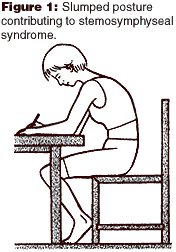
While the goal of neurodevelopment of the locomotor system is to achieve the upright posture, Brugger and Janda have shown how deleterious sedentarism is.2,4,6 Brugger describes the typical sedentary posture of man via a linkage system.6,8 He has shown how approximation of the sternum and symphysis increases both end-range joint loading and muscular tension (see Figure 1). It is possible, however, to demonstrate that postural correction can immediately improve joint function (by centrating joints) and reduce muscle tone.

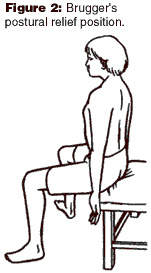
Brugger's postural relief position facilitates muscles, which promotes dynamic stability and reciprocally inhibits muscles, which shorten due to postural stress (see Figures 2 and 3).9 Check tension in the upper trapezius in the slump posture and then in the postural relief position. Muscle tension should be dramatically reduced in the postural relief position. Another check is to have your patient perform cervical rotation in the slump and corrected postures. Again, a dramatic improvement in the postural relief position should be observed.


Take-home message: Brugger's advice is an excellent tool for balancing muscles. It can improve patient compliance with home exercises and increase patient awareness of postural corrections.
Manual Resistance Techniques for Restoring Muscle Balance
During development of upright posture, the infant gradually learns to use various points of support by substituting dynamic stabilization muscle activity for tonic reflex muscle activity. This dynamic stabilization function is first observed around one month of age as the infant begins to orient its head to visual or auditory stimuli.
As the infant develops its motor skill and is gradually able to verticalize its neck, then lumbar spine and extremities, tonic muscle activity is replaced by coactivation of antagonists. Short cervical extensors along with deep neck flexors allow the head to be raised, with the cervicocranial junction held in a stable neutral or centrated posture. Development from one stage to another requires that balanced muscle contraction of antagonists replaces dominance of tonic muscular activity. This coactivation centrates or aligns joints in maximum congruence. This coactivation is not reflex (brain stem) but supraspinal, and is the beginning point of postural motor activity in the human.
In the upper extremities, tonic activity (flexion, internal rotation, adduction and pronation) is joined with dynamic stabilization activity (extension, external rotation, abduction and supination). This allows the arms to be moved from a sucking position to reaching, grasping and locomotion positions. In the lower extremities, tonic activity (ankle plantar flexion and inversion, hip flexion, internal rotation and adduction) is joined with dynamic stabilization activity (ankle dorsiflexion and eversion, hip external rotation and abduction). This allows the legs to move from a fetal position to one capable of creeping, crawling and (later) kneeling.
Muscle imbalances once formed are easily perpetuated by reciprocal inhibition. The tighter muscles continuously inhibit their weakened antagonists, thus perpetuating the problem. Additionally, it is the overactive muscles, which are regularly trained by most exercise routines unless specific attention is paid to isolating the inhibited "weak link." Janda has always maintained that tight or overactive muscles should be relaxed or stretched before initiating a strengthening routine or else the muscle imbalance will only be reinforced.10 Stabilization exercise programs place a great emphasis on conscious control of posture during exercise so as to carefully isolate the inhibited muscles and thus avoid encouraging inappropriate muscle substitution and faulty movement patterns.11,12
Another method which Brugger has developed is to strongly facilitate by eccentric contraction the inhibited muscles of the upper and lower extremity. This manual resistance technique (MRT) takes very little time, yet achieves a powerful reciprocal inhibition of the entire chain of overactive muscles. This eccentric MRT is performed in the upper quarter by resisting finger abduction; finger and wrist extension; supination; and shoulder external rotation and abduction. In the lower quarter, eccentric MRT is performed by resisting ankle eversion and dorsiflexion; hip abduction; and finally, hip external rotation.
Only three or four repetitions of each MRT are necessary. Pre- and post-checks of adverse tension, such as trigger points in the upper trapezius, pectoralis major or thigh adductors, or straight leg raising or wrist extension mobility, should be performed to confirm the positive effect.
Take-home message: Eccentric MRTs applied to key inhibited muscles of the extremities are a quick way to reciprocally inhibit postural muscle hypertonia.
Conclusion
The concept of muscle imbalance is reinforced by knowledge of neurodevelopment of the upright posture. Chains form in our patients, which are invaluable aids in troubleshooting. It is not enough to simply identify a muscle imbalance and treat those muscles. The chain that is dysfunctional must be identified, and treatment of a key link given. Supraspinal control, which begins after three weeks of life, is the beginning of voluntary motor control. If chains of agonist-antagonist muscle incoordination hypothetically related to various stages of neurodevelopment of the upright posture are improved, this may be a significant advance in our treatment of motor system problems. Research into motor system problems. Research into agonist-antagonist coactivation, joint congruence, equilibrium, maximization of joint load handling ability and neurological programs in the adult representative of neurodevelopmental stages is eagerly anticipated.
References
- Janda V. On the concept of postural muscles and posture in man. Aus J Physioth 1983;29:83-84.
- Liebenson C (ed.) Rehabilitation of the Spine: A Practitioner's Manual. Baltimore: Williams and Wilkins, 1995.
- Liebenson CS. Active muscular relaxation techniques, part two: clinical application. JMPT 1990;13(1).
- Lewit K. Manipulative Therapy in Rehabilitation of the Motor System, 3rd ed. London: Butterworths, 1999.
- Kolar P. The sensomotor nature of postural functions. Its fundamental role in rehabilitation of the motor system. J of Orthopaedic Medicine 2000 (in sched).
- Lewit K. Chain reactions in the locomotor system in the light of coactivation patterns based on developmental neurology. J of Orthopaedic Medicine 2000 (in sched).
- Lewit K, Kolah P. Chain reactions related to the cervical spine. In: Murphy D. Conservative Management of Cervical Spine Syndromes. Appleton and Lange, 1999.
- Liebenson C. Self-help advice - Brugger relief position. J Bodywork and Movement Therapies 1999;3.
- Liebenson CS, Perri M, Murphy D, DeFranca C. Safe Back Workout: Flexibility, Breathing and Relaxation Routine. Williams and Wilkins, 1998.
- Jull G, Janda V. Muscles and motor control in low back pain. In: Twomney LT, Taylor JR (eds.) Physical Therapy for the Low Back, Clinics in Physical Therapy. New York; Churchill Livingstone, 1987.
- Morgan D. Concepts in functional training and postural stabilization for the low-back injured. Top Acute Care Trauma Rehabil 1988;2(4):8-17.
- Richardson CA, Jull GA. Muscle control - pain control. What exercises would you prescribe? Man Ther 1995;1(1):2-10.


