How Microcurrent Stimulation Produces ATP -- One Mechanism
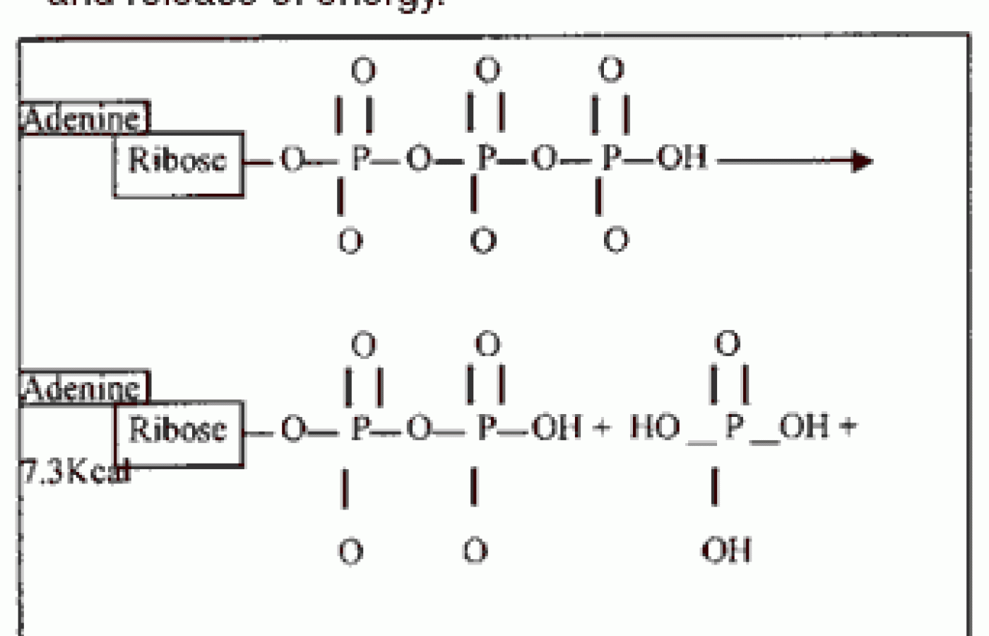
ATP (adenosine triphosphate) molecules are the storage and distribution vehicles for energy in the body. The breakdown of ATP into ADP yields energy. It is the cleaving of the phosphate bond that yields the energy. This energy is utilized in almost all energy related cellular reactions. In addition to being integral to the function of virtually every cell in our body, we may also look at ATP function by categories of activity. Such essential functions include: 1) muscle contraction; 2) protein biosynthesis; 3) nerve transmission; and 4) active transport across cell membranes.
In muscle contraction, the process occurs as such: each muscle spindle is composed of muscle fibers. Inside the muscle fibers are many muscle fibrils. These muscle fibrils are suspended in a fluid matrix called sarcoplasm. Suspended in the sarcoplasm are thousands and thousands of mitochondria, which contain large amounts of ATP.
It is ATP that energizes the muscle contraction process by the ATPase activity of the exposed myosin head. When ATP is exposed to the myosin head, it is cleaved and energy is released. It should be noted that along with ATP, magnesium is very necessary in ATP energy releasing reactions. Before ATP can become "active ATP," magnesium must bind between the second and third phosphate. Clinically, magnesium deficiency may be related to such conditions as fibromyalgia and chronic fatigue syndrome.
Synthesis of almost any chemical compound requires energy. That energy is ATP, which is critically important to the biosynthesis of proteins, phospholipids, purines, pyrimidines and hundreds (if not thousands) of other substances. We will take ATP involvement in protein synthesis as a case in point: a single protein may be composed of many thousands of amino acids. It takes the breakdown of four high- energy phosphate bonds to link two amino acids together.
Maximally, two ATP could serve as energy to join two amino acids together, so if our protein is composed of 10,000 amino acids, it may take 20,000 ATP to form just this one protein. It should also be noted that the amino acids themselves utilize ATP indirectly as they are first co-transported into the cells.
ATP is necessary for nerve transmission. Nerve transmission entails the release of nerve transmitter substance from the presynaptic terminal into the synaptic cleft, which simply put is a space between one nerve and another. The nerve transmitter substance spans the cleft and attaches to the receptor of the other cell. The nerve transmitter substance must be constantly formed anew in the presynaptic terminal for future release; the energy for this formation is supplied by ATP. There are many mitochondria in the presynaptic terminal to form and store the ATP for this process. The formation of ATP will be discussed later in relation to the stimulatory effects of microcurrent.
At the post-synaptic terminal, the next nerve cell down the line, it is through active transport of sodium, potassium and calcium that concentration differences across the nerve cell membrane cause nerve firing and propagation of nerve signals to travel to the next presynaptic terminal. These concentration gradients could not be accomplished without ATPase active transport across nerve cell membranes.
Active transport is brought about by the energy release of ATP in the breakdown of its phosphate bonds (see Figure 1 for ATP chemical structure and energy releasing breakdown).
Active transport is a means of getting molecules across the cell membrane, either into or out of the cell, against a concentration gradient. That concentration gradient may be electrical or a pressure gradient. Sodium, potassium, calcium, glucose, amino acids and many other compounds are transported this way.
To summarize, ATP is the energy currency for our bodies. In reality, virtually every cytological, histological and physiological process is ATP-mediated, which makes ATP clinically important. While our bodies in theory can produce all the ATP we need, the fact is that it doesn't. Microcurrent stimulation between 200-800 microamps is a way of supercharging the tissue with ATP, which will reside there until needed. By this means, much of the research that shows a 200% increase inhealing rate can be explained as it applies to hundreds of conditions. In a clinical sense, any healing process takes a great deal of ATP and may be accelerated through a means of increasing ATP in the tissue. Microcurrent stimulation accomplishes this by increasing ATP in the tissue by up to 400%.
Discussion
Microcurrent stimulation to the body causes radically increased production of ATP levels. This allows the body to perform whatever healing process it has undertaken in an accelerated fashion. It may even allow one to get over the proverbial "hump" that was unachievable, due to insufficient ATP concentrations to perform the changes needed.
ATP is the dynamic reservoir of energy in our body. Glucose serves as a more long-term reservoir but in itself does relatively little to fuel the body. Glucose is first converted to ATP. ATP is the storage and distribution vehicle for energy. From the moment an ATP molecule is produced, it is typically consumed within one minute.
The turnover rate for ATP is very high. However, the body does have a vast capacity to store ATP. One can build ATP reserves. This is one reason that, unlike other forms of electric therapy such as interferential, or higher amperage TENS and galvanic, microcurrent stands unique in that it has a cumulative effect, rather than a diminishing effect. Other electric stimulation devices decrease ATP levels.
Moreover, these devices cannot even be thought of in the realms of ATP generation. It has been shown that any stimulation over 1,000 microamps causes plateauing and then reduction in ATP. Microcurrent therapy, which is used from one to usually 600 uA clinically, is the modality of choice for increased tissue healing. Research and clinical trials have shown that with microcurrent stimulation, there is a 40-50% reduction in healing time of ulcers and sprain/strains; fractures heal faster and stronger; and that even bad scarring (keloid scars) remodel to become a healthier, stronger scar. Other ATP related microcurrent stimulatory effects include decreased inflammation, edema and swelling, and increased physical endurance in sports.
Clinically, microcurrent stimulation is not at all limited to its effects of increased ATP production in its capacity as a treatment modality, but its effects to reduce injury healing time in half are truly dramatic. The mechanism for increased ATP production from microcurrent electric stimulation can be explained. In the following text, its mechanism, from the grosser external application down to the molecular level, is revealed.
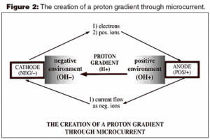
A microcurrent instrument delivers a direct current (DC), and so the nature of its electrical flow is the same. Figure 2 is a representation of a typical circuit in which electrons flow from the cathode to the anode, while current, in the form of negative ions, flows from the anode to the cathode. The negative ions can be thought of as an offset to the electrons which flow the other way. This comprises the circuit.
Of more clinical relevance is what takes place at the anode and the cathode rather than the circuit itself. Around the cathode, which is negative, is an environment of negative hydroxyl ions (OH-). This is caused by electron interaction with water molecules at the cathode, hydrolyzing the water molecules into hydrogen and hydroxyl molecules. The same reaction takes place at the anode; however, because the anode is positive in polarity, protons (hydrogen ions) form the environment around the anode.
In the instantaneous moment, both hydrogen and hydroxyl ions are forms around both pad electrodes. However, over time, the ionic environment becomes hydrogen around the anode electrode and hydroxyl around the cathode electrode. Because it is hydrogen that leads to the creation of ATP, it then follows that as a residual effect after the microcurrent stimulator is turned off, ATP production continues at the site.
Meanwhile, at the negative electrode, ATP production ceases immediately at the turning off of the stimulation because there is no residual hydrogen cloud in the area. Protons (H+) produce a very powerful effect here. Looking at Figure 2, we see that the protons diffuse to an area of less protons, namely from the anode side towards the cathode side. As the protons (H+) migrate through the tissue, they cause increased formation of ATP.
This ATP formation can be explained through the chemosmotic theory of Mitchell. This theory explains how mitochondria form ATP by familiar processes such as the electron transport chain and Krebs cycle. In Mitchell's theory, we see that ionized hydrogen (protons) trigger the electron transport chain by combining with NADH to form NADH+, as well as FAD to form FADH2 and other mediators. The net effect of each cycle of the electron transport chain is the introduction of six hydrogen ions inbetween the inner and outer mitochondrial membranes.
At this point, hydrogen ATPase is activated by the high intramembrane content of hydrogen and activates ATP production. This is accomplished by the addition of a phosphate group to ADP to form ATP (see Figure 1, except note that the process will basically run in reverse to form ATP). This process is known as oxidative phosphorylation. The ATP is at this point transferred out of the mitochondria into the cytoplasm of the cell where it is stored until utilization.
Conclusion and Afterthoughts
ATP can be produced by the body by many means other than those mentioned above. However, it is a very dynamic energy source, and at the site of injury or at a site of overuse and microinjury, ATP supplies can become diminished.
Microcurrent therapy offers a unique and wonderful answer to tissue healing. Clinically, microcurrent therapy is also a therapy of choice for hyperacute injuries in that it produces little if any sensation. It should also be remembered or known that in addition to its uniqueness, microcurrent therapy shares many of the qualities of other electric therapies: namely for application in pain control, muscle relaxation and nerve re-education. It also stands somewhat unique in its ability to increase vascular permeability and in its use as a means of electroacupuncture. MENS microcurrent instruments have the ability to detect the bioelectric state of the human body, and have proven a standard of technical excellence unsurpassed in clinical modality.
References
Guyton AC, Hall JE. Textbook of Medical Physiology. W.B. Saunders Company.
Insel PM. Perspectives in Nutrition. Mosby.
Kroschwitz JI, Winokur M. Chemistry - General, Organic, Biological. McGraw-Hill, Inc.
Giancoli DC. Physics: Principles with Applications. Prentice Hall.
Cheng, et al. The effects of electric current on ATP generation, protein synthesis, and membrane transport in rat skin. Clinical Orthopaedics and Related Research Nov/Dec 1982;171.
Carley and Wainapel. Electrotherapy for acceleration of wound healing: low intensity direct current. Archives of Physical Medicine and Rehabilitation July 1985; vol. 66.
Becker RO. Cross Currents: The Perils of Electropollution, the Promise of Electromedicine. G.P. Putnam's Sons.
In Appreciation of: Dan Worley; Dr. Dennis & Carolyn Greenlee; Dr. Thomas Wing; and Monad Corporation.
Building on a historic March 2026 meeting between Make America Healthy Again and chiropractic leadership, MAHA has announced the launch of the MAHA Chiropractic Hub, “a coordinated national partnership uniting MAHA Center, MAHA Action, and the chiropractic profession, including national associations, state organizations, practitioners, educators, researchers, and patient advocates. The Chiropractic Hub will advance federal policy, expand patient access, and build broad public support for chiropractic care across America.”
The chiropractic profession is confronting one of the most significant federal regulatory threats it has faced in decades. A new U.S. Department of Education (ED) accountability framework – commonly referred to as the “Do No Harm” (DNH) regulation – could place many chiropractic programs at risk of losing access to federal financial aid (student loans), potentially reshaping the future of chiropractic education and workforce development across the United States.
Pain has become the dominant language of musculoskeletal healthcare. Numeric pain-rating scales and symptom reports are routinely used as primary indicators of clinical success. But while pain reduction is meaningful, it is an incomplete and often misleading representation of recovery. This has real consequences for patient adherence, long-term outcomes, and how conservative care is perceived within the broader healthcare system.