New York's highest court of appeals has held that no-fault insurers cannot deny no-fault benefits where they unilaterally determine that a provider has committed misconduct based upon alleged fraudulent conduct. The Court held that this authority belongs solely to state regulators, specifically New York's Board of Regents, which oversees professional licensing and discipline. This follows a similar recent ruling in Florida reported in this publication.
The Forward Head Posture
In our last article, we discussed posture in general; ways to differentiate good versus bad posture; and some of the physiological effects of altered posture.1 We used an image of forward head posture to exemplify a type of bad posture and also discussed some of the related stresses, strains and physiological changes that will impact cervical tissues. The focus of this article, forward head posture, will examine methods of postural assessment; skeletal alignment position on radiographs; research efforts to determine normal lateral cervical posture; and clinical interventions for forward head posture.
Forward head posture is a clinical entity that has been identified by multiple authors as a significant factor in a variety of musculoskeletal pain syndromes.2-7 Although some reports are essentially anecdotal,2-4 several reports use sophisticated statistical analyses and healthy controls versus painful subjects to establish forward head posture as a real clinical entity with significant musculoskeletal consequences.5-7
Unfortunately, the assessment of head posture in relation to the thorax and the subsequent assessment of the underlying skeletal geometry are subjects that have been largely neglected in chiropractic training. We will discuss the assessment of forward head posture and review a couple of methods devised by both chiropractic and PT innovators for the reduction of this common postural displacement.
Postural Assessment
Normal postural alignment of the head over the thorax in the lateral view has been described as the vertical alignment of the external auditory meatus over the acromioclavicular joint.3,4,8 This position can be assessed easily, with the use of a plumb line with the patient in the relaxed neutral position. To document the existence of forward head posture syndrome, some examiners will employ the use of a Polaroid camera mounted to a leveled tripod and loaded with special film that has gridlines applied to the film.
Fortunately, a number of authors have assessed the repeatability of the relaxed neutral position or "self-balance position"9 with the use of sophisticated biplanar photography,10 lateral cephalometric radiographs9 and lateral cervical radiographs.11 In each case, subjects were instructed to assume a neutral relaxed posture prior to assessment.
In one study, subjects were instructed to tilt the head forward and backward with decreasing amplitude until they reached the most relaxed position. They were then instructed to look into the eyes of their own reflected image in a mirror placed two meters away.9 In another study, the subjects were instructed to simply close their eyes, flex, extend the head twice, come to their most neutral relaxed position, hold this position still, and then open their eyes.11
In all of these studies, the test subjects returned to the same self-balance position when tested over a variety of time periods: one minute,10 two minutes,10 one hour,9 one week10 and 12 weeks.11 These results established the repeatability of the self-balance position as a stable posture over time. Using these methods, or slightly modified versions of these methods, has resulted in the establishment of the forward head posture as a condition with significant musculoskeletal consequences of increased pain and alterations in overall spinal mobility.5-7
Skeletal Alignment Position
At least two simple methods exist that determine various spinal and head positions on lateral radiographs. The first was developed by dental and orthodontia researchers to measure the relative positions of the skull and axis vertebra. An additional measurement was made by comparing true vertical and horizontal lines on lateral cephalometric radiographs to a line connecting the posterior tip of the odontiod process of C2 and the posteroinferior body margin of C4.9 Although highly reliable and reproducible, this method has less relevance to chiropractic practice than the next method described.
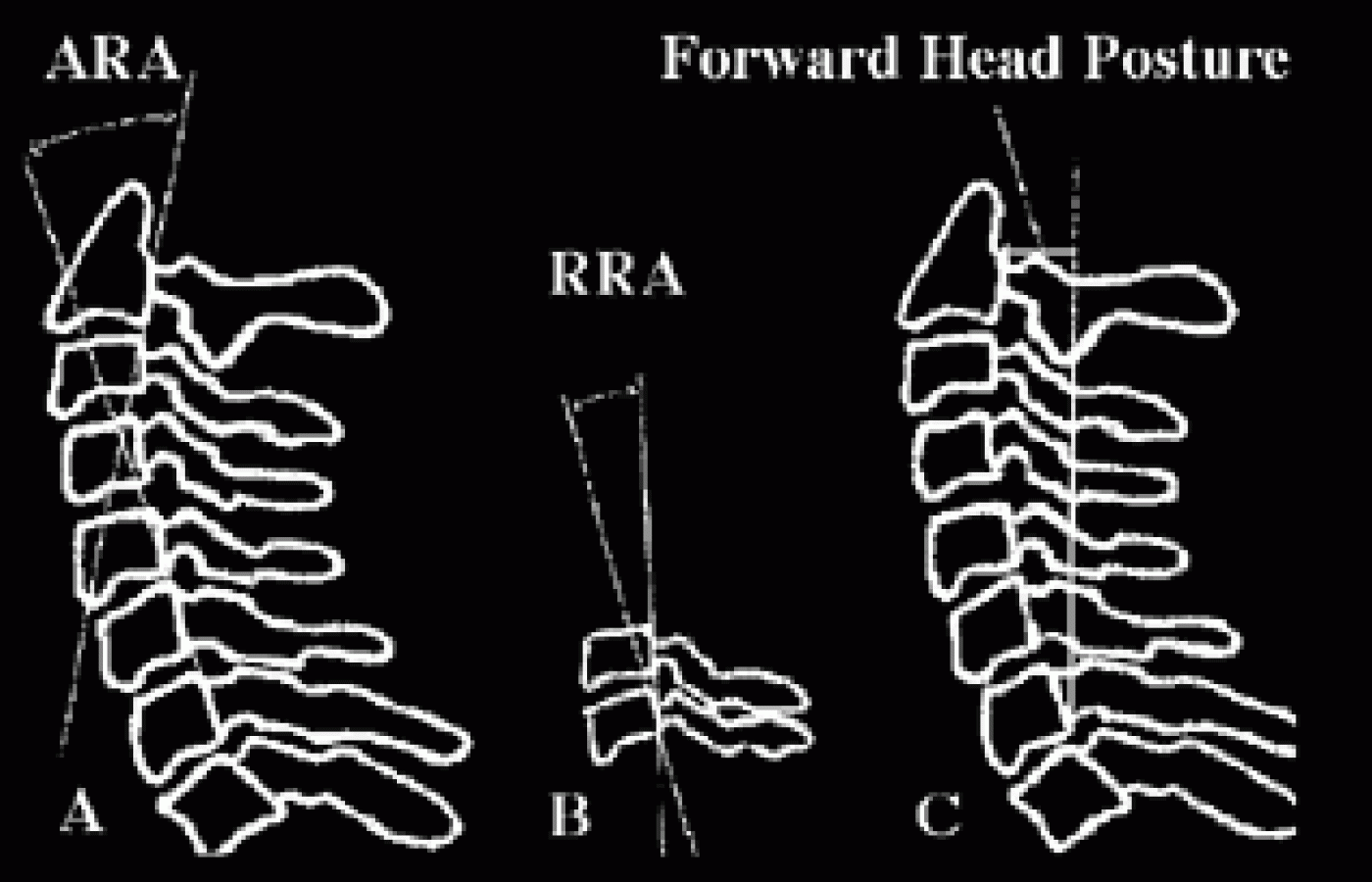
Jackson et al.12 describe a method of determining the relative position of adjacent vertebrae on neutral lateral cervical radiographs by constructing lines drawn across the posterior vertebral body margins. They termed the measurement of the angle of intersection of such lines relative rotation angles. Overall cervical lordosis is determined by measuring the angle of intersection between lines drawn across the posterior vertebral body margins of C2 and C7. Jackson et al. termed this measure of overall cervical lordosis as the absolute rotation angle of the cervical spine. Finally, Jackson et al. describe a method of measuring forward head posture on lateral cervical radiographs. They constructed a superior vertical line from the posteroinferior corner of C7 and measured the perpendicular distance from this line to the posterosuperior portion of the vertebral body of C2. These measures are illustrated in Figure 1 and represent a repeatable and reliable method to quantify the cervical lordosis, and any amount of forward head posture that exists in your patients.

Figure 1: A) Absolute rotation angle (ARA) of overall lordosis magnitude. B) Relative rotation angle (RRA) or magnitude of intersegmental angle. C) Linear measure of forward displacement of head relative to upper dorsal spine.
What Is Normal?
As already described above, normal postural alignment when performing visual inspection exists when the external auditory meatus lines up directly over the acromioclavicular joint. Several authors have described on lateral cervical radiographs the attributes of normal spinal geometry in patients who have no history of neck pain,13 and in patients without cervicocranial symptoms who were selected using specified biomechanical criteria.14
Gore et al.13 described the geometric configuration of the cervical spine in 200 asymptomatic people between the ages of 20 and 60. When comparing the angle of intersection of lines drawn along the posterior vertebral body margins of C2 and C7 (as described above), they found the average degree of overall cervical lordosis was 21 degrees. Similarly, Harrison et al.14 found the overall average degree of lordosis between C2 and C7 in their asymptomatic subjects was 34 degrees.
This establishes a normal average range of lordosis between C2 and C7 of 21-34 degrees in so-called "normal subjects." When assessing the magnitude of forward head posture, Harrison et al.14 found an average of about 15 millimeters of forward displacement of the head in relation to the thorax, using the measurement described above and depicted in Figure 1C (see above).
Finally, Harrison et al.14 found the average range of relative rotation angles for the lower cervical spine (i.e., C3-C4 through C6-C7) to vary between 6.26 and 7.18 degrees for the geometric position of adjacent vertebrae. The average relative rotation angle found for C2-C3 was 7.59 degrees and was explained as being larger overall in comparison to the other relative rotation angles because C2 is a larger vertebrae and would naturally make up a larger portion of a circular lordosis in the cervical spine. These values can now serve as standards against which to compare patients and to assess outcome for interventions designed to reduce the clinical entity of the forward head posture.
Clinical Interventions for the Forward Head Posture
Two studies demonstrate interventions that can easily be provided by chiropractors that have been shown to reduce the magnitude of the forward head posture. Pearson and Walmsley15 described their findings in terms of resting posture with a group of 30 subjects who performed three sets of 10 repetitions of neck retraction exercises. In this exercise, subjects are instructed to pull "... the head and neck posteriorly into a position in which the head is aligned more directly over the thorax, while the head and eyes remain level."15 They found that after performing the second set of exercises, the subjects' neutral resting posture demonstrated a statistically significant (p<0.05) reduction in forward head posture that averaged approximately 4mm.
Additionally, Harrison et al.11 found an average overall improvement in cervical lordosis of 13.2 degrees measured between C2 and C7, and an average of 9.8mm reduction in forward head posture following a regimen of 12 weeks of maximum tolerance cervical extension traction in a retrospective sample of 35 subjects. These results were compared to a control group that received no intervention over a similar 12-week period and whose cervical alignment and posture showed no change.
These two studies, although preliminary in nature, demonstrate two methods that chiropractors can routinely use in their efforts to reduce the effects of the forward head posture syndrome. One method is "passive" (extension traction), while the other involves active participation of the patient (exercise). One can only speculate what synergistic effect these two methods might have if used together. One of us (ST) routinely uses these two methods in clinical practice and empirically has observed consistent results in the reduction of the forward head posture with loss of or reduction in cervical lordosis. A controlled clinical trial would be necessary to confirm this clinical observation.
Conclusion
The forward head posture is an abnormality of posture routinely observed in subjects with a wide variety of musculoskeletal complaints. Both medical and chiropractic researchers have devised reliable methods to assess the posture and skeletal alignment of the spine and skull of patients with such abnormalities. Methods which have been shown to reduce the magnitude of the forward head posture are readily available to doctors of chiropractic. For those interested in these methods, we would suggest investigating both the McKenzie protocols of neck retraction exercise15 and extension traction protocols.11
References
- Seaman D, Troyanovich S. The chasm between posture and chiropractic education and treatment. Dyn Chiro 2000;18(1):20-22.
- Mennell JM. The Musculoskeletal System: Differential Diagnosis from Symptoms and Physical Signs. Gaithersburg, Maryland: Aspen Publishers, Inc., 1992, pp. 126-33.
- Donatelli R, Wooden M. Orthopaedic Physical Therapy. New York: Churchill Livingstone Inc., 1989.
- Cailliet R. Soft Tissue Pain and Disability. Philadelphia: FA Davis Co., 1977.
- Haughie LJ, Fiebert IM, Roach KE. Relationship of forward head posture and cervical backward bending to neck pain. J Manual Manipulative Ther 1995;3:91-7.
- Greenfield B, Catlin PA, Coats PW, et al. Posture in patients with shoulder overuse injuries and healthy individuals. JOSPT 1995;21:287-95.
- Griegel-Morris P, Larson K, Mueller-Klaus K, Oatis CA. Incidence of common postural abnormalities in the cervical shoulder, and thoracic regions and their association with pain in two age groups of healthy subjects. Phys Ther 1992;72:425-31.
- Lee D. Principles and practices of muscle energy and functional techniques. In: Grieve GP (ed.) Modern Manual Therapy of the Vertebral Column. New York: Churchill Livingstone, 1986.
- Sandham A. Repeatability of head posture recordings from lateral cephalometric radiographs. British J Orthodontics 1988;15:157-62.
- Refshauge K, Goodsell M, Lee M. Consistency of cervical and cervicothoracic posture in standing. Australian Physiotherapy 1994;40;235-9.
- Harrison DD, Jackson BL, Troyanovich S, et al. The efficacy of cervical extension-compression traction combined with diversified manipulation and drop table adjustments in the rehabilitation of cervical lordosis: a pilot study. J Manipulative Physiol Ther 1994;17:454-64.
- Jackson BL, Harrison DD, Robertson GA, Barker WF. Chiropractic biophysics lateral cervical film analysis reliability. J Manipulative Physiol Ther 1993;16:384-91.
- Gore DR, Sepic SB, Gardner GM. Roentgenographic findings of the cervical spine in asymptomatic people. Spine 1986;6:521-4.
- Harrison DD, Janik TJ, Troyanovich SJ, Holland B. Comparisons of lordotic cervical spine curvatures to a theoretical ideal model of the static sagittal cervical spine. Spine 1996;21:667-75.
- Pearson ND, Walmsley RP. Trial into the effects of repeated neck retractions in normal subjects. Spine 1995;20:1245-51.


