It’s a new year and many chiropractors are evaluating what will enhance their respective practices, particularly as it relates to their bottom line. One of the most common questions I get is: “Do I need to be credentialed to bill insurance, and what are the best plans to join?” It’s a loaded question – but one every DC ponders. Whether you're already in-network or pondering whether to join, here's what you need to know.
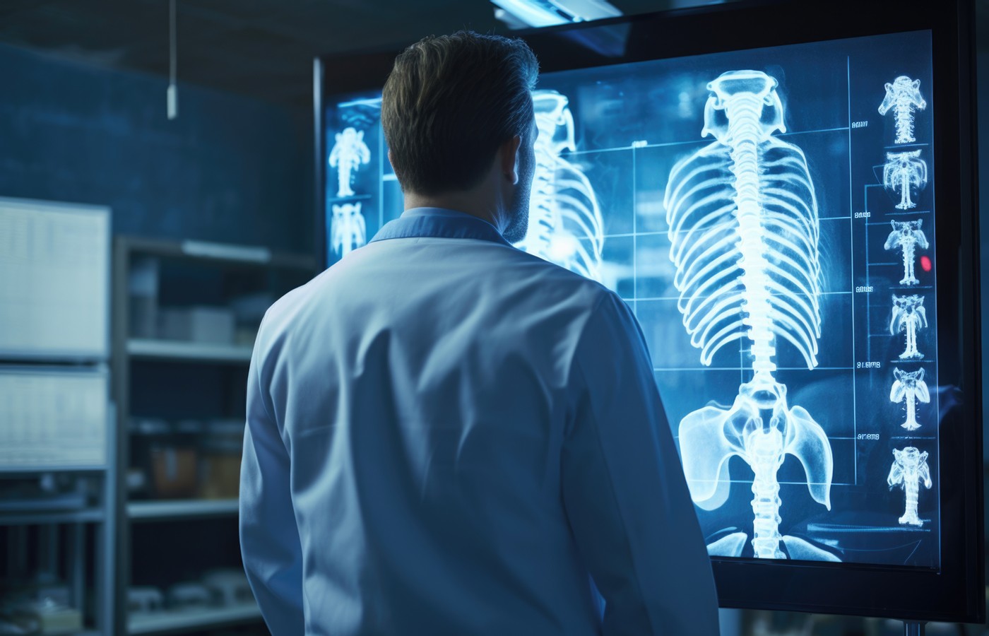
When Is Radiographic Imaging Medically Necessary for LBP?
- It has been reported that diagnostic imaging (on initial evaluation) does not usually provide a benefit to patients with LBP, and most clinical practice guidelines (CPGs) do not recommend it unless there are red flags.
- This article reviews a putative patient presentation with acute low back pain to determine medical necessity for imaging.
- This patient responded well to care with only two spinal manipulations and trigger-point treatments. There was no need for imaging of his spine.
As a student at Logan College of Chiropractic from 1968-1972, we were taught to X-ray every new patient. While serving in chiropractic clerkship in the college clinic, we were required to use the Gonstead marking system. The purpose was focused on biomechanics. The most important reasons were to reveal spinal scoliosis and spinal subluxations.
We were also taught that we might discover a pathology with the use of radiographic studies. And of course, it was commonly believed that X-ray examinations would reduce the potential for a malpractice case.
While working at the Lovelace Medical Center in Albuquerque, N.M., one of the primary care physicians stated that imaging every patient prior to receiving chiropractic was not medically necessary. It was at that point I reconsidered the need for the imaging of every patient to avoid malpractice.
It became obvious to me that the use of spinal manipulation did not require spinal imaging, especially with a 14x36-inch radiographic image. Currently, evidence-based practice does not support the use of routine radiographic examinations in order to support the use of spinal manipulation.
Inappropriate diagnostic imaging contributes to the increasing costs of spinal health care and may expose patients to unnecessary radiation, depending on the diagnostic imaging procedure performed. It has been reported that diagnostic imaging (on initial evaluation) does not usually provide a benefit to patients with LBP, and most clinical practice guidelines (CPGs) do not recommend it unless there are red flags.1
Evidence-based guidelines recommend that radiographic examination of low back pain patients requires the presence of “red flags.” Clinical Compass guidelines clarify the red flags that demonstrate the need for imaging the low back pain patient:2
- A history and physical exam to detect red flags (underlying serious pathologies), signs of nerve compression and/or injuries (fracture, dislocation, etc.)
- A consideration of X-rays if the patient experienced blunt trauma or if there is no response to treatment after 4-6 weeks of conservative care
- Urgent specialized imaging for back and neck pain with critical qualities: sphincter or gait disturbance, saddle anesthesia, severe or progressive neurologic deficit, systemic illness (cancer, infection), vascular causes (suspected abdominal/thoracic aorta aneurysm), or cervical artery dissection
A Case Example
With the foundation for the use of spinal imaging and medical necessity documented, let’s review a putative patient presentation with acute low back pain.
Subjective Examination
Chief concern: “My back is killing me.” This 35-year-old male presents with acute low back pain that started three days prior to the visit. He does not know why his back hurts and denies any particular injury. He has been treated successfully for acute low back pain episodes by other chiropractic physicians over the past 10 years. “Usually, I get relief with one or two adjustments.”
The low back pain first bothered him following a lifting injury at work 12 years ago. He mentions that he has been under more stress at work, which requires an increase of time at his desk and computer.
Although he denies smoking, he is drinking more alcohol and consuming more food. Eating and drinking reduce his stress.
He started running to reduce his weight and stress, but the low back ache became a sharp pain on the right side of his lower back. He tried to run through the pain, but then noticed spasms in the back, which interfered with his sleep. Hot baths do reduce the low back ache.
He denies any radiations and points to the area of the lumbosacral spine on the right as the area of pain. Today, he rates his severity at 5/10 because the pain is bothering him at work. He claims this time the pain is worse than previous episodes.
Objective Examination
This middle-aged mesomorphic male appears mildly overweight and in distress.
Vital signs: height 5’10”, weight 225 lbs., B/P 140/86, pulse 73.
Posture: inferior and posterior left iliac crest and anterior and superior right iliac crest.
Gillet test: positive for fixation of the left iliac joint.
Palpation: reproduces the CC pain at L5/S1 right and over the supraspinous ligaments.
Myofascial trigger points: palpation reveals taut bands and painful nodules in the multifidi muscles on the right at L4-S1 right, and the left iliopsoas muscle.
Long sit test: demonstrates functional leg-length inequality on the left.
Kemp’s maneuver: negative for leg pain, but produces localized pain at the right L5/S1 joint with reduced range of motion in right lateral and extension positions.
Straight-leg raise: right lower extremity produces pain at the right L5/S1 joint at 95 degrees. The left SLR is without pain at 100 degrees.
Posterior joint dysfunction at L5/S1 with pain, reduced ROM, and hypertonicity of the paravertebral muscles.
Sensory intact for the L4-5-S1 dermatomes (sharp/dull) bilaterally.
Motor intact with 5/5 L4-5-S1 myotomes bilaterally.
Deep tendon reflexes (myotatic) 2+ bilaterally for the L4-5-S1 reflexes.
Babinski sign absent bilaterally.
Assessment / Plan
Assessment:
- Acute low back pain due to stress
- Lumbar facet syndrome
Treatment Plan:
- Spinal manipulation to reduce pain and improve spinal joint function
- Trigger-point pressure release to reduce pelvic obliquity and functional leg-length inequality of the left lower extremity
- Provide 2-3 treatments with follow-up evaluation on each visit
- RX weight loss and a regular exercise program to reduce spinal stress
- Consider an imaging study if the patient does not respond to care within two weeks
Outcomes / Discussion
This patient responded well to care with only two spinal manipulations and trigger-point treatments. There was no need for imaging of his spine. That said, had he not responded to care and the symptoms were progressively worsening, possibly with radiations into the lower extremities, I would have performed another physical examination and considered imaging to evaluate for degenerative joint and disc disease.
Quiz Time
1. Spinal imaging would have been indicated had he demonstrated which one of the following symptoms?
a. Good response to care
b. Radiating pain down the extremities and saddle anesthesia
c. Aching pain following the initial spinal manipulation
d. None of the above
2. What is the rationale for treating the left iliopsoas muscle?
a. To reduce the pelvic obliquity and functional leg-length inequality
b. To correct anatomical leg-length inequality
c. To reduce the lumbar facet syndrome
d. None of the above
Quiz Answers: 1. B. 2. A.
References
- Taylor DN, Hawk C. An investigation into chiropractic intern adherence to radiographic guidelines in clinical decisions with a descriptive comparison to clinical practitioners. J Chiropr Educ, 2023 Mar 1;37(1):41-49.
- Diagnostic Imaging. Clinical Compass; available at https://clinicalcompass.org/evidence-center-research-summaries/diagnostic-imaging/.


