It’s a new year and many chiropractors are evaluating what will enhance their respective practices, particularly as it relates to their bottom line. One of the most common questions I get is: “Do I need to be credentialed to bill insurance, and what are the best plans to join?” It’s a loaded question – but one every DC ponders. Whether you're already in-network or pondering whether to join, here's what you need to know.
Chest Study vs. Thoracic Study

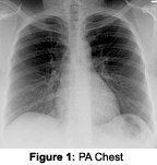
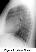
There are significant technical differences between a thoracic spine series and a chest series. The clinical reasons for performing these studies often overlap but should not be viewed as commensurate studies. A chest series is performed mainly for evaluating possible pathology involving the lungs, heart and mediastinum - all soft-tissue structures. A thoracic spine series is performed to evaluate possible pathology involving the thoracic spine and paraspinal structures - all mainly osseous or cartilaginous structures.


The lateral chest view is taken with a non-grid technique, using a 14 x 17 cassette at 72" film focal distance at a kVp of 100 to 110, with a time commensurate with patient's thickness. Collimation is again a little less than the film size and, in general, there is no need to change the position of the cassette from the PA view. For both views, the central ray is at about T6. In general, the left side always is the side closest to the cassette. The X-ray is again taken on inspiration.
The routine thoracic spine study includes the following: AP and lateral thoracic spine views. The AP thoracic view is taken with a grid technique, using a 7 x 17 or 14 x 17, at 40" film focal distance and a kVp of 76-80, with a time commensurate with patient's thickness. Collimation is a little less than the 7 x 17 cassette. If possible, filter the top 1/3 of the film or use the anode heel effect. Central ray is about the level of T6. X-ray is done on inspiration.

Obviously, there is a significant difference between a chest study and a thoracic spine study. Note the higher kVp and lower time for the chest study and the lower kVp and higher time for thoracic spine study. Remember that the high-kV/low-mAs techniques produce a wide gray scale, and low-kV/high-mAs techniques produce a higher-contrast X-ray. Pathologies can be missed very easily if the wrong technique is used or if one wished to "cover all the bases." It is not possible to obtain a diagnostic-quality AP thoracic/chest view on one film. If the clinical symptoms make it difficult to determine whether or not there is lung or thoracic spine pathology, perform a routine thoracic study and a PA chest.
For more specific technical information on performing chest or T-spine series, the following resources will be very helpful:
- Jaeger S. Atlas of Radiographic Positioning. Appleton & Lange, 1988.
- Vlasuk S. Tips on thoracic spine technique. Available at: www.drvxray.com.
Your chiropractic college radiology departments always are accessible for questions on X-ray technique. Your local DACBR is a great source for assistance on X-ray technique and diagnostic review. For information on most DACBRs in the U.S. and internationally, visit www.accr.org.


