Because they have yet to pass national legislation protecting the chiropractic profession, Japanese DCs are in a similar situation that U.S. DCs faced. We were fortunate enough to be able to pass chiropractic licensure state by state. The DCs in Japan must accomplish this nationally, which has proved to be an extremely difficult task. And in spite of their efforts, Japanese DCs are currently faced with two chiropractic professions.
Normal vs. Abnormal Gas in the Abdomen
Lumbar spine and pelvic plain-film radiographic exams commonly are performed in chiropractic offices. Obviously, these are ordered primarily to evaluate the bony structures. The soft tissues, however, also are visible and deserve our attention, as they can frequently give us clues to disease processes that may be present. If we are concerned with the soft tissue of the abdomen, specific abdominal views can be performed. This article will review the normal and abnormal gas patterns that can be seen in the abdomen.
Normal Gas in the Abdomen
Gas is frequently seen throughout the gastrointestinal tract. In most cases, it is of no clinical significance. The majority of this gas originates as swallowed air. Other sources include carbonated beverages and gas-forming bacteria within the colon.
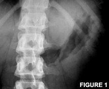
Gas commonly is seen in the stomach on an upright lumbar film. This gas is located in the fundus of the stomach and often will be part of an air-fluid interface. Occasionally, the rugal folds of the stomach are visible. These are oriented to the long axis of the stomach and have a wavy appearance. (See Figure 1.) The composition of gas in the stomach is very similar to that of the air we breathe. The gastric fundus is fixed in location and typically measures within 2.5 cm of the left hemidiaphragm.
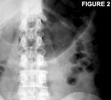
The large bowel typically contains more gas than the small bowel. This is due to decreased absorption in addition to the actual production of gas by bacteria. The composition of the gas changes due to this bacterial production. It frequently will have hydrogen, carbon dioxide and methane added to the nitrogen. The large bowel is identified by the gas-filled haustral outpouchings. Each haustral outpouching is bordered by a plica semilunaris, a fold of colonic wall that projects into the lumen. The plica semilunaris extend only partially around the circumference of the bowel, as opposed to the valvulae conniventes of the small bowel. Gas in the large bowel frequently is accompanied by fecal material. Small gas pockets frequently are trapped within the fecal material, giving it a characteristic mottled appearance. Large-bowel gas is more likely to be located around the periphery of the abdomen. But like the small bowel, it can have a wide variation in its location. The only fixed regions are the hepatic and splenic flexures.
Abnormal Gas in the Abdomen
Occasionally, a mass can be seen deviating gas collections in the stomach. This is a difficult diagnosis to make accurately on plain films, due to the fact that the entire lumen of the stomach often is not outlined by the gas and the presence of swallowed liquids. Narrowing of the stomach lumen also might be visible. This often is due to scirrhous carcinoma or lymphoma. Again, this is difficult to see on a single plain film and usually requires multiple plain films over time, showing persistent narrowing.
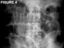
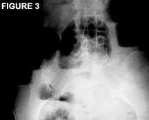
Three or more air fluid levels in the small bowel are considered abnormal. (See Figure 3.) This is a result of diminished/lost bowel transport. So, an obstruction or a loss/diminished peristalsis would be the cause. Small-bowel loops should be less than 3 cm in diameter. Small-bowel distention is considered present if the loops are greater than 3 cm in diameter. Figure 4 demonstrates small-bowel distention. Note the central location of the gas and the visible valvulae conniventes, helping to identify this as the small bowel. Some of the possible causes of small-bowel distention are mechanical obstruction, volvulus, intussusception, hernias and postoperative.
Small-bowel dilation can uncommonly reach and exceed 5cm in diameter and be mistaken for large-intestine dilatation. Occasionally, an isolated loop of distended small bowel can be visualized. This is referred to as a sentinel loop and is due to an inflammatory process near the loop, such as acute pancreatitis. Within the small bowel, a linear collection of gas bubbles is known as the "string of pearls" sign and is an indicator of mechanical small-bowel obstruction. The actual site of small-bowel obstruction may be visualized by an abrupt ending or tapering of a dilated loop of the small bowel. Often times, the actual site of obstruction is not known with just a plain-film study.
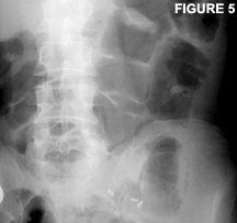
Any air fluid levels in the large bowel are considered abnormal. Just as described in the small bowel, this indicates a diminished/lost bowel transport, either due to obstruction or a loss/diminished peristalsis. Distention of the large bowel often is readily visualized on plain films. Distention generally is considered present if the diameter of the large bowel is greater than 5 cm. Figure 5 demonstrates dilatation of the transverse and descending colon. Masses and strictures also might be suspected on plain-film examination. Again, due to the variable appearance of large-intestine gas and the presence of fecal material, these are all difficult diagnoses to make accurately off plain-film studies. If masses or strictures are suspected, further imaging such as an abdominal CT may be warranted. Causes of large-bowel distention include mechanical obstruction, volvulus, adynamic ileus, toxic megacolon and ischemic colitis.
Gas outside the confines of the GI tract always is considered abnormal. There are several radiographic signs of free gas in the peritoneum, called pneumoperitoneum. The first is the double-wall sign or bas-relief sign. Both sides of the bowel wall are visible, due to intraluminal and extraluminal gas. The bowel wall will stand out and be readily visible when this happens. Figure 6 demonstrates this concept. The next sign of pneumoperitoneum is called the football sign and is typically only seen in children. It is a large collection of gas in the anterior peritoneum, vertically orientated in the shape of a football. Another sign of pneumoperitoneum is free air outlining the undersurface of the diaphragms. Gas commonly is seen below the left hemidiaphragm, but should clearly be associated with the gastric fundus or splenic flexure of the colon. The last sign of pneumoperitoneum is called the falciform ligament sign. The free gas in the abdomen will outline the water-dense falciform ligament, which can be seen in the right-upper medial abdomen.

Branching gas patterns occasionally can be seen in the region of the liver. These may be due to gas in the biliary tree or in the portal venous system. If the branching gas pattern is seen toward the periphery of the liver, it is indicative of gas within the portal veins. This is an ominous sign and may indicate mesenteric infarction. If the branching gas pattern is seen toward the central region of the liver, it is indicative of gas within the biliary tree. This is not as ominous and may be associated with things such as gallstone ileus, duodenal ulcer perforation and postoperative status.
Conclusion
Since the abdominal soft tissues are visible when performing lumbar spine or pelvic plain-film exam, an understanding of the normal gas patterns of the abdomen is important, so that abnormal patterns may be identified.
References
- Baker SR. The Abdominal Plain Film, First Edition. Norwalk, Conn.: Appleton & Lange, 1990.
- Burgener F, Kormano M. Differential Diagnosis in Conventional Radiology, First Edition. New York: Thieme-Stratton, Inc, 1985.
- Chapman S, Nakielny R. Aids to Radiological Differential Diagnosis, Fourth Edition. New York: WB Saunders, 2003.


